Improving performance of CNN to predict likelihood of COVID-19 using chest X-ray images with preprocessing algorithms
Paper and Code
Jun 11, 2020


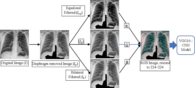
As the rapid spread of coronavirus disease (COVID-19) worldwide, chest X-ray radiography has also been used to detect COVID-19 infected pneumonia and assess its severity or monitor its prognosis in the hospitals due to its low cost, low radiation dose, and wide accessibility. However, how to more accurately and efficiently detect COVID-19 infected pneumonia and distinguish it from other community-acquired pneumonia remains a challenge. In order to address this challenge, we in this study develop and test a new computer-aided diagnosis (CAD) scheme. It includes several image pre-processing algorithms to remove diaphragms, normalize image contrast-to-noise ratio, and generate three input images, then links to a transfer learning based convolutional neural network (a VGG16 based CNN model) to classify chest X-ray images into three classes of COVID-19 infected pneumonia, other community-acquired pneumonia and normal (non-pneumonia) cases. To this purpose, a publicly available dataset of 8,474 chest X-ray images is used, which includes 415 confirmed COVID-19 infected pneumonia, 5,179 community-acquired pneumonia, and 2,880 non-pneumonia cases. The dataset is divided into two subsets with 90% and 10% of images in each subset to train and test the CNN-based CAD scheme. The testing results achieve 94.0% of overall accuracy in classifying three classes and 98.6% accuracy in detecting Covid-19 infected cases. Thus, the study demonstrates the feasibility of developing a CAD scheme of chest X-ray images and providing radiologists useful decision-making supporting tools in detecting and diagnosis of COVID-19 infected pneumonia.
 Add to Chrome
 Add to Firefox
 Add to Edge