Yusaku Hayashi
Plaque segmentation via masking of the artery wall
Jan 26, 2022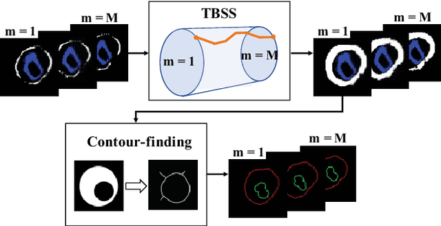
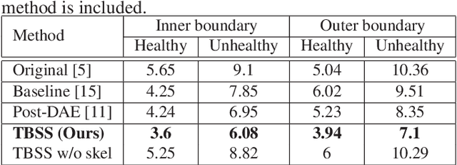

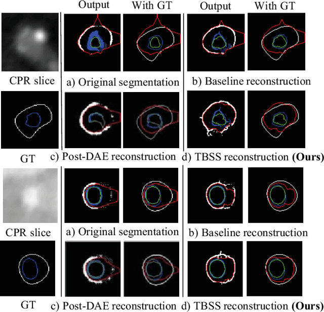
Abstract:The presence of plaques in the coronary arteries are a major risk to the patients' life. In particular, non-calcified plaques pose a great challenge, as they are harder to detect and more likely to rupture than calcified plaques. While current deep learning techniques allow precise segmentation of regular images, the performance in medical images is still low, caused mostly by blurriness and ambiguous voxel intensities of unrelated parts that fall on the same range. In this paper, we propose a novel methodology for segmenting calcified and non-calcified plaques in CCTA-CPR scans of coronary arteries. The input slices are masked so only the voxels within the wall vessel are considered for segmentation. We also provide an exhaustive evaluation by applying different types of masks, in order to validate the potential of vessel masking for plaque segmentation. Our methodology results in a prominent boost in segmentation performance, in both quantitative and qualitative evaluation, achieving accurate plaque shapes even for the challenging non-calcified plaques. We believe our findings can lead the future research for high-performance plaque segmentation.
Coronary Wall Segmentation in CCTA Scans via a Hybrid Net with Contours Regularization
Feb 27, 2020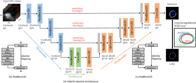
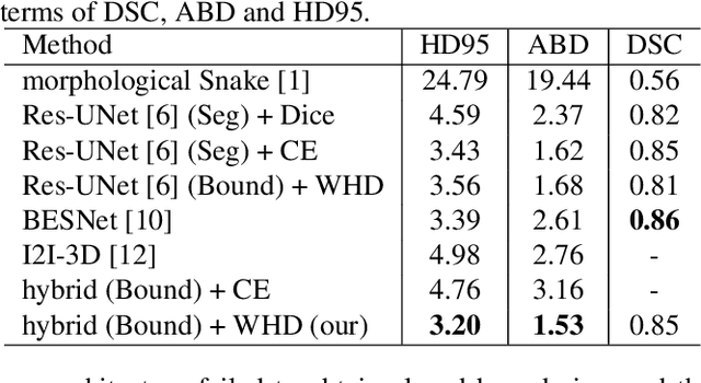

Abstract:Providing closed and well-connected boundaries of coronary artery is essential to assist cardiologists in the diagnosis of coronary artery disease (CAD). Recently, several deep learning-based methods have been proposed for boundary detection and segmentation in a medical image. However, when applied to coronary wall detection, they tend to produce disconnected and inaccurate boundaries. In this paper, we propose a novel boundary detection method for coronary arteries that focuses on the continuity and connectivity of the boundaries. In order to model the spatial continuity of consecutive images, our hybrid architecture takes a volume (i.e., a segment of the coronary artery) as input and detects the boundary of the target slice (i.e., the central slice of the segment). Then, to ensure closed boundaries, we propose a contour-constrained weighted Hausdorff distance loss. We evaluate our method on a dataset of 34 patients of coronary CT angiography scans with curved planar reconstruction (CCTA-CPR) of the arteries (i.e., cross-sections). Experiment results show that our method can produce smooth closed boundaries outperforming the state-of-the-art accuracy.
 Add to Chrome
 Add to Firefox
 Add to Edge