Takashi Azuma
Textile-based conformable and breathable ultrasound imaging probe
Nov 07, 2023Abstract:Daily monitoring of internal tissues with conformable and breathable ultrasound (US) imaging probes is promising for early detection of diseases. In recent years, textile substrates are widely used for wearable devices since they satisfy both conformability and breathability. However, it is not currently possible to use textile substrates for US probes due to the reflection or attenuation of US waves at the air gaps in the textiles. In this paper, we fabricated a conformable and breathable US imaging probe by sandwiching the US elements between two woven polyester textiles on which copper electrodes were formed through electroless plating. The air gaps between the fibers at the electrode parts were filled with copper, allowing for high penetration of US waves. On the other hand, the non-electrode parts retain air gaps, leading to high breathability. The fabricated textile-based probe showed low flexural rigidity ($0.066 \times 10^{-4} N \cdot m^2/m$) and high air permeability ($11.7 cm^3 / cm^2 \cdot s$). Human neck imaging demonstrated the ability of the probe to monitor the pulsation of the common carotid artery and change in the internal jugular vein diameter, which lead to the early detection of health issues such as arteriosclerosis and dehydration.
Blind Signal Separation for Fast Ultrasound Computed Tomography
Apr 27, 2023


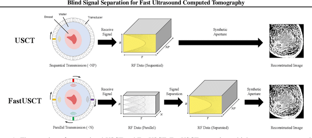
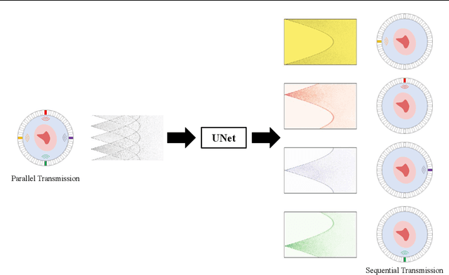
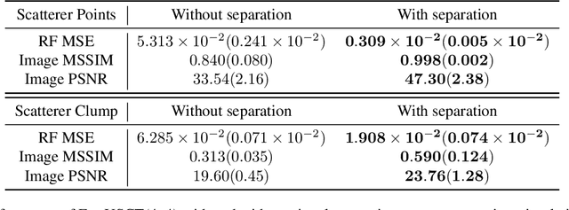
Abstract:Breast cancer is the most prevalent cancer with a high mortality rate in women over the age of 40. Many studies have shown that the detection of cancer at earlier stages significantly reduces patients' mortality and morbidity rages. Ultrasound computer tomography (USCT) is considered as a promising screening tool for diagnosing early-stage breast cancer as it is cost-effective and produces 3D images without radiation exposure. However, USCT is not a popular choice mainly due to its prolonged imaging time. USCT is time-consuming because it needs to transmit a number of ultrasound waves and record them one by one to acquire a high-quality image. We propose FastUSCT, a method to acquire a high-quality image faster than traditional methods for USCT. FastUSCT consists of three steps. First, it transmits multiple ultrasound waves at the same time to reduce the imaging time. Second, it separates the overlapping waves recorded by the receiving elements into each wave with UNet. Finally, it reconstructs an ultrasound image with a synthetic aperture method using the separated waves. We evaluated FastUSCT on simulation on breast digital phantoms. We trained the UNet on simulation using natural images and transferred the model for the breast digital phantoms. The empirical result shows that FastUSCT significantly improves the quality of the image under the same imaging time to the conventional USCT method, especially when the imaging time is limited.
 Add to Chrome
 Add to Firefox
 Add to Edge