Stephen M. Plaza
Fully-Automatic Synapse Prediction and Validation on a Large Data Set
Apr 11, 2016


Abstract:Extracting a connectome from an electron microscopy (EM) data set requires identification of neurons and determination of synapses between neurons. As manual extraction of this information is very time-consuming, there has been extensive research effort to automatically segment the neurons to help guide and eventually replace manual tracing. Until recently, there has been comparatively less research on automatically detecting the actual synapses between neurons. This discrepancy can, in part, be attributed to several factors: obtaining neuronal shapes is a prerequisite first step in extracting a connectome, manual tracing is much more time-consuming than annotating synapses, and neuronal contact area can be used as a proxy for synapses in determining connections. However, recent research has demonstrated that contact area alone is not a sufficient predictor of synaptic connection. Moreover, as segmentation has improved, we have observed that synapse annotation is consuming a more significant fraction of overall reconstruction time. This ratio will only get worse as segmentation improves, gating overall possible speed-up. Therefore, we address this problem by developing algorithms that automatically detect pre-synaptic neurons and their post-synaptic partners. In particular, pre-synaptic structures are detected using a Deep and Wide Multiscale Recursive Network, and post-synaptic partners are detected using a MLP with features conditioned on the local segmentation. This work is novel because it requires minimal amount of training, leverages advances in image segmentation directly, and provides a complete solution for polyadic synapse detection. We further introduce novel metrics to evaluate our algorithm on connectomes of meaningful size. These metrics demonstrate that complete automatic prediction can be used to effectively characterize most connectivity correctly.
Large-Scale Electron Microscopy Image Segmentation in Spark
Apr 01, 2016


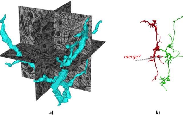
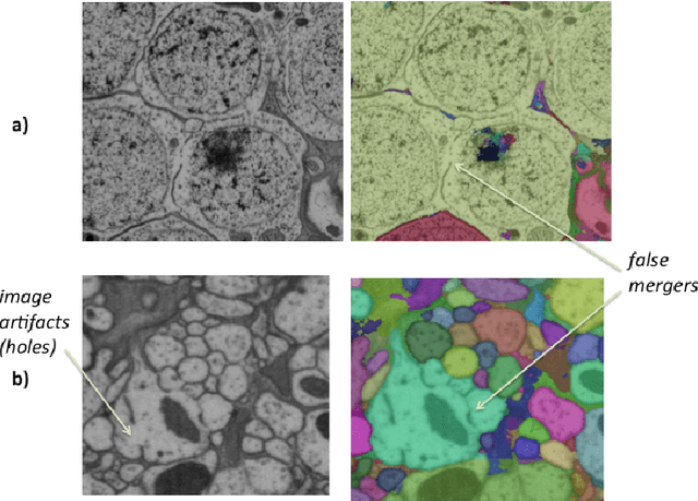
Abstract:The emerging field of connectomics aims to unlock the mysteries of the brain by understanding the connectivity between neurons. To map this connectivity, we acquire thousands of electron microscopy (EM) images with nanometer-scale resolution. After aligning these images, the resulting dataset has the potential to reveal the shapes of neurons and the synaptic connections between them. However, imaging the brain of even a tiny organism like the fruit fly yields terabytes of data. It can take years of manual effort to examine such image volumes and trace their neuronal connections. One solution is to apply image segmentation algorithms to help automate the tracing tasks. In this paper, we propose a novel strategy to apply such segmentation on very large datasets that exceed the capacity of a single machine. Our solution is robust to potential segmentation errors which could otherwise severely compromise the quality of the overall segmentation, for example those due to poor classifier generalizability or anomalies in the image dataset. We implement our algorithms in a Spark application which minimizes disk I/O, and apply them to a few large EM datasets, revealing both their effectiveness and scalability. We hope this work will encourage external contributions to EM segmentation by providing 1) a flexible plugin architecture that deploys easily on different cluster environments and 2) an in-memory representation of segmentation that could be conducive to new advances.
Annotating Synapses in Large EM Datasets
Dec 04, 2014


Abstract:Reconstructing neuronal circuits at the level of synapses is a central problem in neuroscience and becoming a focus of the emerging field of connectomics. To date, electron microscopy (EM) is the most proven technique for identifying and quantifying synaptic connections. As advances in EM make acquiring larger datasets possible, subsequent manual synapse identification ({\em i.e.}, proofreading) for deciphering a connectome becomes a major time bottleneck. Here we introduce a large-scale, high-throughput, and semi-automated methodology to efficiently identify synapses. We successfully applied our methodology to the Drosophila medulla optic lobe, annotating many more synapses than previous connectome efforts. Our approaches are extensible and will make the often complicated process of synapse identification accessible to a wider-community of potential proofreaders.
Focused Proofreading: Efficiently Extracting Connectomes from Segmented EM Images
Sep 03, 2014


Abstract:Identifying complex neural circuitry from electron microscopic (EM) images may help unlock the mysteries of the brain. However, identifying this circuitry requires time-consuming, manual tracing (proofreading) due to the size and intricacy of these image datasets, thus limiting state-of-the-art analysis to very small brain regions. Potential avenues to improve scalability include automatic image segmentation and crowd sourcing, but current efforts have had limited success. In this paper, we propose a new strategy, focused proofreading, that works with automatic segmentation and aims to limit proofreading to the regions of a dataset that are most impactful to the resulting circuit. We then introduce a novel workflow, which exploits biological information such as synapses, and apply it to a large dataset in the fly optic lobe. With our techniques, we achieve significant tracing speedups of 3-5x without sacrificing the quality of the resulting circuit. Furthermore, our methodology makes the task of proofreading much more accessible and hence potentially enhances the effectiveness of crowd sourcing.
 Add to Chrome
 Add to Firefox
 Add to Edge