Shanaka Ramesh Gunasekara
Spatio-Temporal Joint Density Driven Learning for Skeleton-Based Action Recognition
May 29, 2025Abstract:Traditional approaches in unsupervised or self supervised learning for skeleton-based action classification have concentrated predominantly on the dynamic aspects of skeletal sequences. Yet, the intricate interaction between the moving and static elements of the skeleton presents a rarely tapped discriminative potential for action classification. This paper introduces a novel measurement, referred to as spatial-temporal joint density (STJD), to quantify such interaction. Tracking the evolution of this density throughout an action can effectively identify a subset of discriminative moving and/or static joints termed "prime joints" to steer self-supervised learning. A new contrastive learning strategy named STJD-CL is proposed to align the representation of a skeleton sequence with that of its prime joints while simultaneously contrasting the representations of prime and nonprime joints. In addition, a method called STJD-MP is developed by integrating it with a reconstruction-based framework for more effective learning. Experimental evaluations on the NTU RGB+D 60, NTU RGB+D 120, and PKUMMD datasets in various downstream tasks demonstrate that the proposed STJD-CL and STJD-MP improved performance, particularly by 3.5 and 3.6 percentage points over the state-of-the-art contrastive methods on the NTU RGB+D 120 dataset using X-sub and X-set evaluations, respectively.
Joint Temporal Pooling for Improving Skeleton-based Action Recognition
Aug 18, 2024


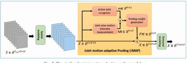
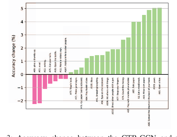
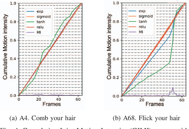
Abstract:In skeleton-based human action recognition, temporal pooling is a critical step for capturing spatiotemporal relationship of joint dynamics. Conventional pooling methods overlook the preservation of motion information and treat each frame equally. However, in an action sequence, only a few segments of frames carry discriminative information related to the action. This paper presents a novel Joint Motion Adaptive Temporal Pooling (JMAP) method for improving skeleton-based action recognition. Two variants of JMAP, frame-wise pooling and joint-wise pooling, are introduced. The efficacy of JMAP has been validated through experiments on the popular NTU RGB+D 120 and PKU-MMD datasets.
A Systematic Approach for MRI Brain Tumor Localization, and Segmentation using Deep Learning and Active Contouring
Feb 06, 2021


Abstract:One of the main requirements of tumor extraction is the annotation and segmentation of tumor boundaries correctly. For this purpose, we present a threefold deep learning architecture. First classifiers are implemented with a deep convolutional neural network(CNN) andsecond a region-based convolutional neural network (R-CNN) is performed on the classified images to localize the tumor regions of interest. As the third and final stage, the concentratedtumor boundary is contoured for the segmentation process by using the Chan-Vesesegmentation algorithm. As the typical edge detection algorithms based on gradients of pixel intensity tend to fail in the medical image segmentation process, an active contour algorithm defined with the level set function is proposed. Specifically, Chan- Vese algorithm was applied to detect the tumor boundaries for the segmentation process. To evaluate the performance of the overall system, Dice Score,Rand Index (RI), Variation of Information (VOI), Global Consistency Error (GCE), Boundary Displacement Error (BDE), Mean absolute error (MAE), and Peak Signal to Noise Ratio (PSNR) werecalculated by comparing the segmented boundary area which is the final output of the proposed, against the demarcations of the subject specialists which is the gold standard. Overall performance of the proposed architecture for both glioma and meningioma segmentation is with average dice score of 0.92, (also, with RI of 0.9936, VOI of 0.0301, GCE of 0.004, BDE of 2.099, PSNR of 77.076 and MAE of 52.946), pointing to high reliability of the proposed architecture.
A Feasibility study for Deep learning based automated brain tumor segmentation using Magnetic Resonance Images
Dec 22, 2020


Abstract:Deep learning algorithms have accounted for the rapid acceleration of research in artificial intelligence in medical image analysis, interpretation, and segmentation with many potential applications across various sub disciplines in medicine. However, only limited number of research which investigates these application scenarios, are deployed into the clinical sector for the evaluation of the real requirement and the practical challenges of the model deployment. In this research, a deep convolutional neural network (CNN) based classification network and Faster RCNN based localization network were developed for brain tumor MR image classification and tumor localization. A typical edge detection algorithm called Prewitt was used for tumor segmentation task, based on the output of the tumor localization. Overall performance of the proposed tumor segmentation architecture, was analyzed using objective quality parameters including Accuracy, Boundary Displacement Error (BDE), Dice score and confidence interval. A subjective quality assessment of the model was conducted based on the Double Stimulus Impairment Scale (DSIS) protocol using the input of medical expertise. It was observed that the confidence level of our segmented output was in a similar range to that of experts. Also, the Neurologists have rated the output of our model as highly accurate segmentation.
 Add to Chrome
 Add to Firefox
 Add to Edge