 Add to Chrome
 Add to Firefox
 Add to Edge
FORCE: Feature-Oriented Representation with Clustering and Explanation
Apr 07, 2025


Learning about underlying patterns in data using latent unobserved structures to improve the accuracy of predictive models has become an active avenue of deep learning research. Most approaches cluster the original features to capture certain latent structures. However, the information gained in the process can often be implicitly derived by sufficiently complex models. Thus, such approaches often provide minimal benefits. We propose a SHAP (Shapley Additive exPlanations) based supervised deep learning framework FORCE which relies on two-stage usage of SHAP values in the neural network architecture, (i) an additional latent feature to guide model training, based on clustering SHAP values, and (ii) initiating an attention mechanism within the architecture using latent information. This approach gives a neural network an indication about the effect of unobserved values that modify feature importance for an observation. The proposed framework is evaluated on three real life datasets. Our results demonstrate that FORCE led to dramatic improvements in overall performance as compared to networks that did not incorporate the latent feature and attention framework (e.g., F1 score for presence of heart disease 0.80 vs 0.72). Using cluster assignments and attention based on SHAP values guides deep learning, enhancing latent pattern learning and overall discriminative capability.
Deep Learning based Framework for Automatic Diagnosis of Glaucoma based on analysis of Focal Notching in the Optic Nerve Head
Dec 10, 2021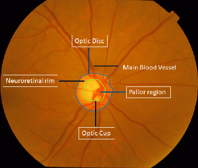
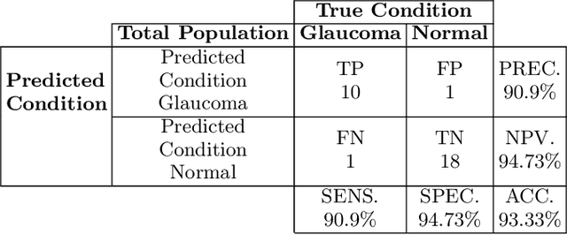
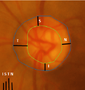
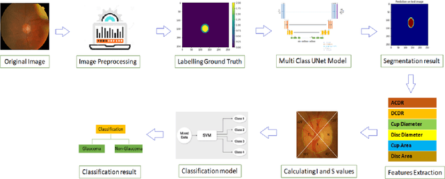
Automatic evaluation of the retinal fundus image is emerging as one of the most important tools for early detection and treatment of progressive eye diseases like Glaucoma. Glaucoma results to a progressive degeneration of vision and is characterized by the deformation of the shape of optic cup and the degeneration of the blood vessels resulting in the formation of a notch along the neuroretinal rim. In this paper, we propose a deep learning-based pipeline for automatic segmentation of optic disc (OD) and optic cup (OC) regions from Digital Fundus Images (DFIs), thereby extracting distinct features necessary for prediction of Glaucoma. This methodology has utilized focal notch analysis of neuroretinal rim along with cup-to-disc ratio values as classifying parameters to enhance the accuracy of Computer-aided design (CAD) systems in analyzing glaucoma. Support Vector-based Machine Learning algorithm is used for classification, which classifies DFIs as Glaucomatous or Normal based on the extracted features. The proposed pipeline was evaluated on the freely available DRISHTI-GS dataset with a resultant accuracy of 93.33% for detecting Glaucoma from DFIs.