Rikathi Pal
A Novel Feature Extraction Model for the Detection of Plant Disease from Leaf Images in Low Computational Devices
Oct 01, 2024Abstract:Diseases in plants cause significant danger to productive and secure agriculture. Plant diseases can be detected early and accurately, reducing crop losses and pesticide use. Traditional methods of plant disease identification, on the other hand, are generally time-consuming and require professional expertise. It would be beneficial to the farmers if they could detect the disease quickly by taking images of the leaf directly. This will be a time-saving process and they can take remedial actions immediately. To achieve this a novel feature extraction approach for detecting tomato plant illnesses from leaf photos using low-cost computing systems such as mobile phones is proposed in this study. The proposed approach integrates various types of Deep Learning techniques to extract robust and discriminative features from leaf images. After the proposed feature extraction comparisons have been made on five cutting-edge deep learning models: AlexNet, ResNet50, VGG16, VGG19, and MobileNet. The dataset contains 10,000 leaf photos from ten classes of tomato illnesses and one class of healthy leaves. Experimental findings demonstrate that AlexNet has an accuracy score of 87%, with the benefit of being quick and lightweight, making it appropriate for use on embedded systems and other low-processing devices like smartphones.
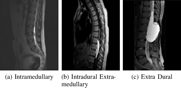
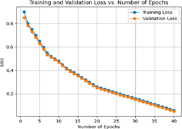
Lumbar Spine Tumor Segmentation and Localization in T2 MRI Images Using AI
May 07, 2024


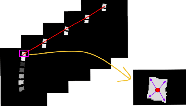
Abstract:In medical imaging, segmentation and localization of spinal tumors in three-dimensional (3D) space pose significant computational challenges, primarily stemming from limited data availability. In response, this study introduces a novel data augmentation technique, aimed at automating spine tumor segmentation and localization through AI approaches. Leveraging a fusion of fuzzy c-means clustering and Random Forest algorithms, the proposed method achieves successful spine tumor segmentation based on predefined masks initially delineated by domain experts in medical imaging. Subsequently, a Convolutional Neural Network (CNN) architecture is employed for tumor classification. Moreover, 3D vertebral segmentation and labeling techniques are used to help pinpoint the exact location of the tumors in the lumbar spine. Results indicate a remarkable performance, with 99% accuracy for tumor segmentation, 98% accuracy for tumor classification, and 99% accuracy for tumor localization achieved with the proposed approach. These metrics surpass the efficacy of existing state-of-the-art techniques, as evidenced by superior Dice Score, Class Accuracy, and Intersection over Union (IOU) on class accuracy metrics. This innovative methodology holds promise for enhancing the diagnostic capabilities in detecting and characterizing spinal tumors, thereby facilitating more effective clinical decision-making.
Panoptic Segmentation and Labelling of Lumbar Spine Vertebrae using Modified Attention Unet
Apr 28, 2024


Abstract:Segmentation and labeling of vertebrae in MRI images of the spine are critical for the diagnosis of illnesses and abnormalities. These steps are indispensable as MRI technology provides detailed information about the tissue structure of the spine. Both supervised and unsupervised segmentation methods exist, yet acquiring sufficient data remains challenging for achieving high accuracy. In this study, we propose an enhancing approach based on modified attention U-Net architecture for panoptic segmentation of 3D sliced MRI data of the lumbar spine. Our method achieves an impressive accuracy of 99.5\% by incorporating novel masking logic, thus significantly advancing the state-of-the-art in vertebral segmentation and labeling. This contributes to more precise and reliable diagnosis and treatment planning.
 Add to Chrome
 Add to Firefox
 Add to Edge