 Add to Chrome
 Add to Firefox
 Add to Edge
Robust Alignment of the Human Embryo in 3D Ultrasound using PCA and an Ensemble of Heuristic, Atlas-based and Learning-based Classifiers Evaluated on the Rotterdam Periconceptional Cohort
Nov 05, 2025Standardized alignment of the embryo in three-dimensional (3D) ultrasound images aids prenatal growth monitoring by facilitating standard plane detection, improving visualization of landmarks and accentuating differences between different scans. In this work, we propose an automated method for standardizing this alignment. Given a segmentation mask of the embryo, Principal Component Analysis (PCA) is applied to the mask extracting the embryo's principal axes, from which four candidate orientations are derived. The candidate in standard orientation is selected using one of three strategies: a heuristic based on Pearson's correlation assessing shape, image matching to an atlas through normalized cross-correlation, and a Random Forest classifier. We tested our method on 2166 images longitudinally acquired 3D ultrasound scans from 1043 pregnancies from the Rotterdam Periconceptional Cohort, ranging from 7+0 to 12+6 weeks of gestational age. In 99.0% of images, PCA correctly extracted the principal axes of the embryo. The correct candidate was selected by the Pearson Heuristic, Atlas-based and Random Forest in 97.4%, 95.8%, and 98.4% of images, respectively. A Majority Vote of these selection methods resulted in an accuracy of 98.5%. The high accuracy of this pipeline enables consistent embryonic alignment in the first trimester, enabling scalable analysis in both clinical and research settings. The code is publicly available at: https://gitlab.com/radiology/prenatal-image-analysis/pca-3d-alignment.
* Submitted version of paper accepted at International Workshop on Preterm, Perinatal and Paediatric Image Analysis 2025
The 4D Human Embryonic Brain Atlas: spatiotemporal atlas generation for rapid anatomical changes using first-trimester ultrasound from the Rotterdam Periconceptional Cohort
Mar 10, 2025Early brain development is crucial for lifelong neurodevelopmental health. However, current clinical practice offers limited knowledge of normal embryonic brain anatomy on ultrasound, despite the brain undergoing rapid changes within the time-span of days. To provide detailed insights into normal brain development and identify deviations, we created the 4D Human Embryonic Brain Atlas using a deep learning-based approach for groupwise registration and spatiotemporal atlas generation. Our method introduced a time-dependent initial atlas and penalized deviations from it, ensuring age-specific anatomy was maintained throughout rapid development. The atlas was generated and validated using 831 3D ultrasound images from 402 subjects in the Rotterdam Periconceptional Cohort, acquired between gestational weeks 8 and 12. We evaluated the effectiveness of our approach with an ablation study, which demonstrated that incorporating a time-dependent initial atlas and penalization produced anatomically accurate results. In contrast, omitting these adaptations led to anatomically incorrect atlas. Visual comparisons with an existing ex-vivo embryo atlas further confirmed the anatomical accuracy of our atlas. In conclusion, the proposed method successfully captures the rapid anotomical development of the embryonic brain. The resulting 4D Human Embryonic Brain Atlas provides a unique insights into this crucial early life period and holds the potential for improving the detection, prevention, and treatment of prenatal neurodevelopmental disorders.
Towards segmentation and spatial alignment of the human embryonic brain using deep learning for atlas-based registration
May 13, 2020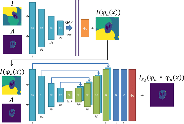
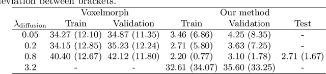

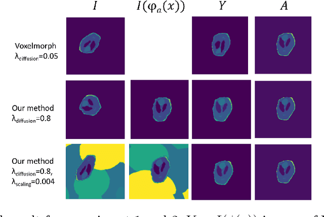
We propose an unsupervised deep learning method for atlas based registration to achieve segmentation and spatial alignment of the embryonic brain in a single framework. Our approach consists of two sequential networks with a specifically designed loss function to address the challenges in 3D first trimester ultrasound. The first part learns the affine transformation and the second part learns the voxelwise nonrigid deformation between the target image and the atlas. We trained this network end-to-end and validated it against a ground truth on synthetic datasets designed to resemble the challenges present in 3D first trimester ultrasound. The method was tested on a dataset of human embryonic ultrasound volumes acquired at 9 weeks gestational age, which showed alignment of the brain in some cases and gave insight in open challenges for the proposed method. We conclude that our method is a promising approach towards fully automated spatial alignment and segmentation of embryonic brains in 3D ultrasound.