Petros-Pavlos Ypsilantis
Learning to detect chest radiographs containing lung nodules using visual attention networks
May 17, 2018


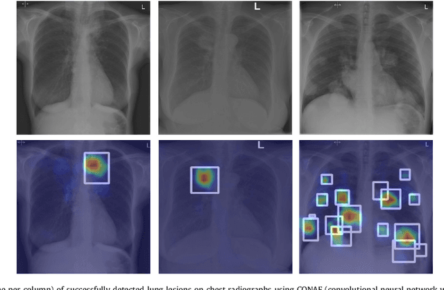
Abstract:Machine learning approaches hold great potential for the automated detection of lung nodules in chest radiographs, but training the algorithms requires vary large amounts of manually annotated images, which are difficult to obtain. Weak labels indicating whether a radiograph is likely to contain pulmonary nodules are typically easier to obtain at scale by parsing historical free-text radiological reports associated to the radiographs. Using a repositotory of over 700,000 chest radiographs, in this study we demonstrate that promising nodule detection performance can be achieved using weak labels through convolutional neural networks for radiograph classification. We propose two network architectures for the classification of images likely to contain pulmonary nodules using both weak labels and manually-delineated bounding boxes, when these are available. Annotated nodules are used at training time to deliver a visual attention mechanism informing the model about its localisation performance. The first architecture extracts saliency maps from high-level convolutional layers and compares the estimated position of a nodule against the ground truth, when this is available. A corresponding localisation error is then back-propagated along with the softmax classification error. The second approach consists of a recurrent attention model that learns to observe a short sequence of smaller image portions through reinforcement learning. When a nodule annotation is available at training time, the reward function is modified accordingly so that exploring portions of the radiographs away from a nodule incurs a larger penalty. Our empirical results demonstrate the potential advantages of these architectures in comparison to competing methodologies.
Learning what to look in chest X-rays with a recurrent visual attention model
Jan 23, 2017


Abstract:X-rays are commonly performed imaging tests that use small amounts of radiation to produce pictures of the organs, tissues, and bones of the body. X-rays of the chest are used to detect abnormalities or diseases of the airways, blood vessels, bones, heart, and lungs. In this work we present a stochastic attention-based model that is capable of learning what regions within a chest X-ray scan should be visually explored in order to conclude that the scan contains a specific radiological abnormality. The proposed model is a recurrent neural network (RNN) that learns to sequentially sample the entire X-ray and focus only on informative areas that are likely to contain the relevant information. We report on experiments carried out with more than $100,000$ X-rays containing enlarged hearts or medical devices. The model has been trained using reinforcement learning methods to learn task-specific policies.
Recurrent Convolutional Networks for Pulmonary Nodule Detection in CT Imaging
Sep 30, 2016


Abstract:Computed tomography (CT) generates a stack of cross-sectional images covering a region of the body. The visual assessment of these images for the identification of potential abnormalities is a challenging and time consuming task due to the large amount of information that needs to be processed. In this article we propose a deep artificial neural network architecture, ReCTnet, for the fully-automated detection of pulmonary nodules in CT scans. The architecture learns to distinguish nodules and normal structures at the pixel level and generates three-dimensional probability maps highlighting areas that are likely to harbour the objects of interest. Convolutional and recurrent layers are combined to learn expressive image representations exploiting the spatial dependencies across axial slices. We demonstrate that leveraging intra-slice dependencies substantially increases the sensitivity to detect pulmonary nodules without inflating the false positive rate. On the publicly available LIDC/IDRI dataset consisting of 1,018 annotated CT scans, ReCTnet reaches a detection sensitivity of 90.5% with an average of 4.5 false positives per scan. Comparisons with a competing multi-channel convolutional neural network for multi-slice segmentation and other published methodologies using the same dataset provide evidence that ReCTnet offers significant performance gains.
 Add to Chrome
 Add to Firefox
 Add to Edge