Oliver Stachs
Visual Analytics for Early Detection of Retinal Diseases
Dec 13, 2022


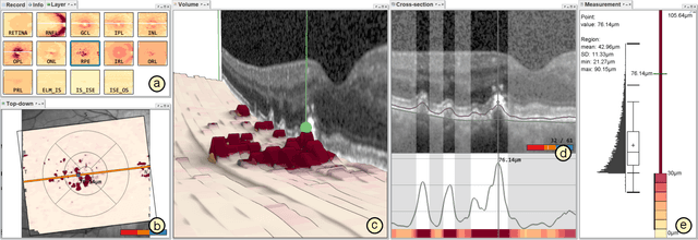
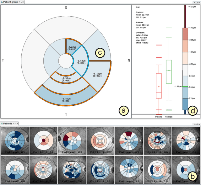
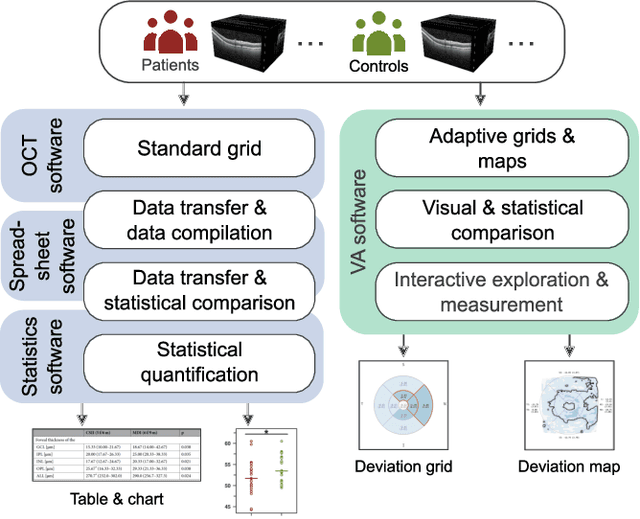
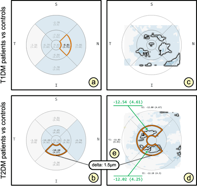
Abstract:Advances in optical coherence tomography (OCT) have enabled noninvasive imaging of substructures of the human retina with high spatial resolution. OCT examinations are now a standard procedure in clinics and an integral part of ophthalmic research. The interpretation of the OCT helps ophthalmologists understand the impact of various retinal and systemic diseases on the structure of the retina in a way not previously possible. In the early stages of retinal diseases, however, the identification and analysis of small and localized substructural changes in the retina remains a challenge. We present an overview of novel visual analytics approaches for the interactive exploration of early retinal changes in single and multiple patients, the comparison of the changes with normative data, and automated quantification and measurement of diagnosis-relevant information. We developed these approaches in close collaboration with ophthalmology researchers and industry experts from a leading OCT device manufacturer. As a result, they not only significantly reduced the time and effort required for OCT data analysis, especially in the context of cross-sectional studies, but have also led to several new discoveries published in biomedical journals.
Transfer Learning with Human Corneal Tissues: An Analysis of Optimal Cut-Off Layer
Jun 22, 2018

Abstract:Transfer learning is a powerful tool to adapt trained neural networks to new tasks. Depending on the similarity of the original task to the new task, the selection of the cut-off layer is critical. For medical applications like tissue classification, the last layers of an object classification network might not be optimal. We found that on real data of human corneal tissues the best feature representation can be found in the middle layers of the Inception-v3 and in the rear layers of the VGG-19 architecture.
 Add to Chrome
 Add to Firefox
 Add to Edge