Moumita Pal
FCM Based Blood Vessel Segmentation Method for Retinal Images
Sep 06, 2012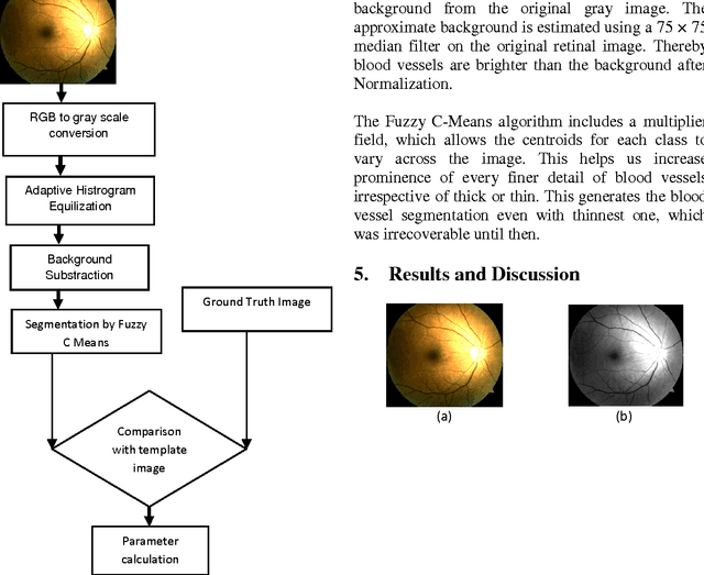
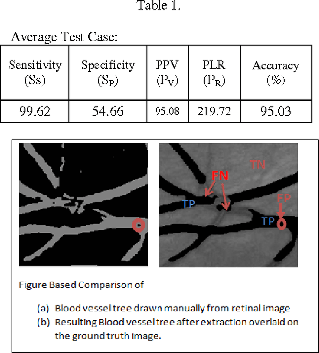
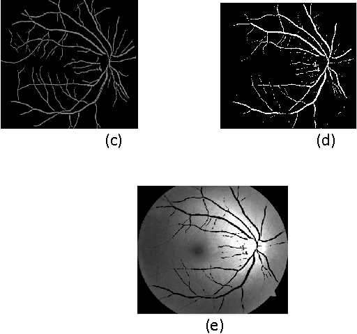
Abstract:Segmentation of blood vessels in retinal images provides early diagnosis of diseases like glaucoma, diabetic retinopathy and macular degeneration. Among these diseases occurrence of Glaucoma is most frequent and has serious ocular consequences that can even lead to blindness, if it is not detected early. The clinical criteria for the diagnosis of glaucoma include intraocular pressure measurement, optic nerve head evaluation, retinal nerve fiber layer and visual field defects. This form of blood vessel segmentation helps in early detection for ophthalmic diseases, and potentially reduces the risk of blindness. The low-contrast images at the retina owing to narrow blood vessels of the retina are difficult to extract. These low contrast images are, however useful in revealing certain systemic diseases. Motivated by the goals of improving detection of such vessels, this present work proposes an algorithm for segmentation of blood vessels and compares the results between expert ophthalmologist hand-drawn ground-truths and segmented image(i.e. the output of the present work).Sensitivity, specificity, positive predictive value (PPV), positive likelihood ratio (PLR) and accuracy are used to evaluate overall performance.It is found that this work segments blood vessels successfully with sensitivity, specificity, PPV, PLR and accuracy of 99.62%, 54.66%, 95.08%, 219.72 and 95.03%, respectively.
* 5 pages,3figures
A Session Based Blind Watermarking Technique within the NROI of Retinal Fundus Images for Authentication Using DWT, Spread Spectrum and Harris Corner Detection
Sep 01, 2012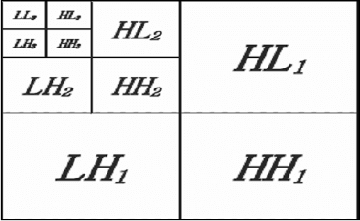
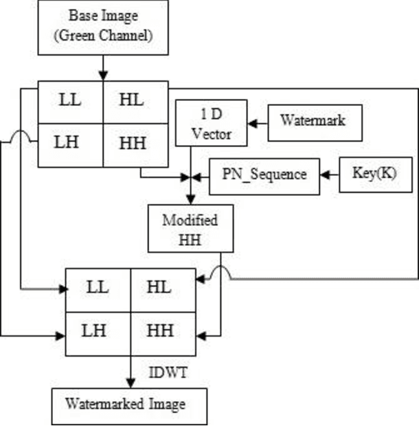

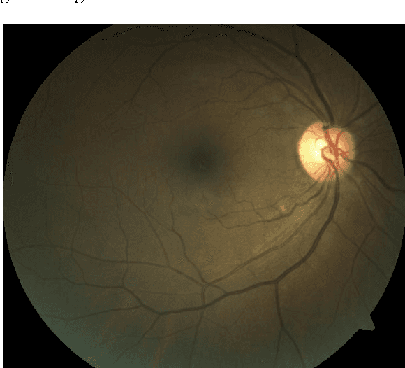
Abstract:Digital Retinal Fundus Images helps to detect various ophthalmic diseases by detecting morphological changes in optical cup, optical disc and macula. Present work proposes a method for the authentication of medical images based on Discrete Wavelet Transformation (DWT) and Spread Spectrum. Proper selection of the Non Region of Interest (NROI) for watermarking is crucial, as the area under concern has to be the least required portion conveying any medical information. Proposed method discusses both the selection of least impact area and the blind watermarking technique. Watermark is embedded within the High-High (HH) sub band. During embedding, watermarked image is dispersed within the band using a pseudo random sequence and a Session key. Watermarked image is extracted using the session key and the size of the image. In this approach the generated watermarked image having an acceptable level of imperceptibility and distortion is compared to the Original retinal image based on Peak Signal to Noise Ratio (PSNR) and correlation value.
* 9 pages, 10 figures
 Add to Chrome
 Add to Firefox
 Add to Edge