Mohamed E. Karar
Multimodal CNN Networks for Brain Tumor Segmentation in MRI: A BraTS 2022 Challenge Solution
Dec 19, 2022


Abstract:Automatic segmentation is essential for the brain tumor diagnosis, disease prognosis, and follow-up therapy of patients with gliomas. Still, accurate detection of gliomas and their sub-regions in multimodal MRI is very challenging due to the variety of scanners and imaging protocols. Over the last years, the BraTS Challenge has provided a large number of multi-institutional MRI scans as a benchmark for glioma segmentation algorithms. This paper describes our contribution to the BraTS 2022 Continuous Evaluation challenge. We propose a new ensemble of multiple deep learning frameworks namely, DeepSeg, nnU-Net, and DeepSCAN for automatic glioma boundaries detection in pre-operative MRI. It is worth noting that our ensemble models took first place in the final evaluation on the BraTS testing dataset with Dice scores of 0.9294, 0.8788, and 0.8803, and Hausdorf distance of 5.23, 13.54, and 12.05, for the whole tumor, tumor core, and enhancing tumor, respectively. Furthermore, the proposed ensemble method ranked first in the final ranking on another unseen test dataset, namely Sub-Saharan Africa dataset, achieving mean Dice scores of 0.9737, 0.9593, and 0.9022, and HD95 of 2.66, 1.72, 3.32 for the whole tumor, tumor core, and enhancing tumor, respectively. The docker image for the winning submission is publicly available at (https://hub.docker.com/r/razeineldin/camed22).
Self-supervised iRegNet for the Registration of Longitudinal Brain MRI of Diffuse Glioma Patients
Nov 20, 2022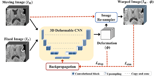
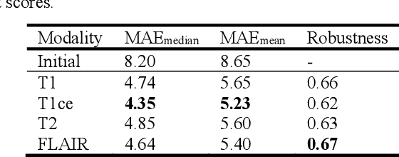
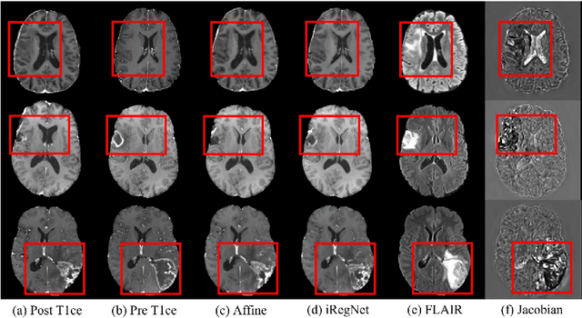
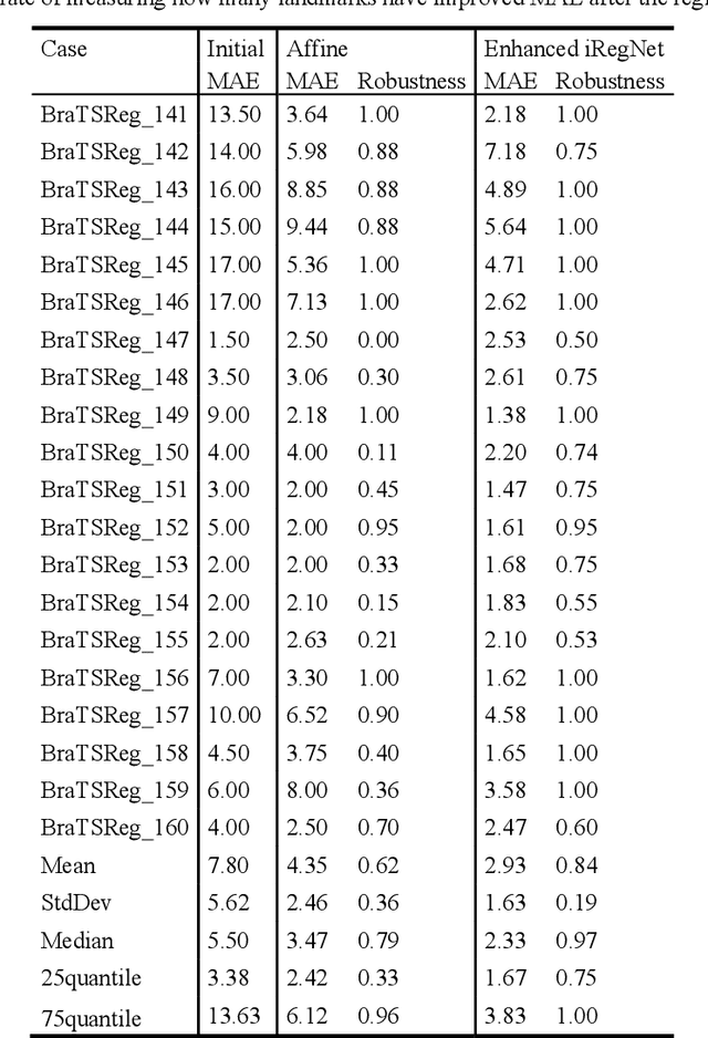
Abstract:Reliable and accurate registration of patient-specific brain magnetic resonance imaging (MRI) scans containing pathologies is challenging due to tissue appearance changes. This paper describes our contribution to the Registration of the longitudinal brain MRI task of the Brain Tumor Sequence Registration Challenge 2022 (BraTS-Reg 2022). We developed an enhanced unsupervised learning-based method that extends the iRegNet. In particular, incorporating an unsupervised learning-based paradigm as well as several minor modifications to the network pipeline, allows the enhanced iRegNet method to achieve respectable results. Experimental findings show that the enhanced self-supervised model is able to improve the initial mean median registration absolute error (MAE) from 8.20 (7.62) mm to the lowest value of 3.51 (3.50) for the training set while achieving an MAE of 2.93 (1.63) mm for the validation set. Additional qualitative validation of this study was conducted through overlaying pre-post MRI pairs before and after the de-formable registration. The proposed method scored 5th place during the testing phase of the MICCAI BraTS-Reg 2022 challenge. The docker image to reproduce our BraTS-Reg submission results will be publicly available.
Ensemble CNN Networks for GBM Tumors Segmentation using Multi-parametric MRI
Dec 27, 2021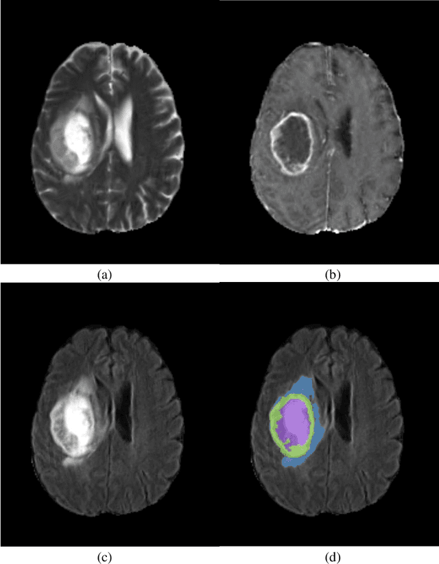

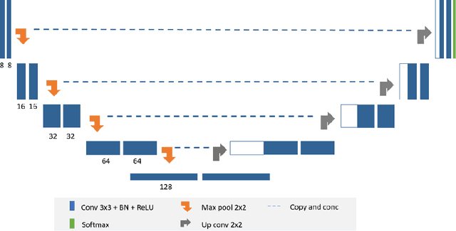
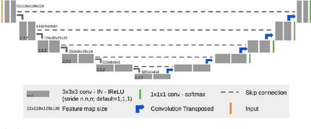
Abstract:Glioblastomas are the most aggressive fast-growing primary brain cancer which originate in the glial cells of the brain. Accurate identification of the malignant brain tumor and its sub-regions is still one of the most challenging problems in medical image segmentation. The Brain Tumor Segmentation Challenge (BraTS) has been a popular benchmark for automatic brain glioblastomas segmentation algorithms since its initiation. In this year, BraTS 2021 challenge provides the largest multi-parametric (mpMRI) dataset of 2,000 pre-operative patients. In this paper, we propose a new aggregation of two deep learning frameworks namely, DeepSeg and nnU-Net for automatic glioblastoma recognition in pre-operative mpMRI. Our ensemble method obtains Dice similarity scores of 92.00, 87.33, and 84.10 and Hausdorff Distances of 3.81, 8.91, and 16.02 for the enhancing tumor, tumor core, and whole tumor regions, respectively, on the BraTS 2021 validation set, ranking us among the top ten teams. These experimental findings provide evidence that it can be readily applied clinically and thereby aiding in the brain cancer prognosis, therapy planning, and therapy response monitoring. A docker image for reproducing our segmentation results is available online at (https://hub.docker.com/r/razeineldin/deepseg21).
DeepSeg: Deep Neural Network Framework for Automatic Brain Tumor Segmentation using Magnetic Resonance FLAIR Images
Apr 26, 2020Abstract:Purpose: Gliomas are the most common and aggressive type of brain tumors due to their infiltrative nature and rapid progression. The process of distinguishing tumor boundaries from healthy cells is still a challenging task in the clinical routine. Fluid-Attenuated Inversion Recovery (FLAIR) MRI modality can provide the physician with information about tumor infiltration. Therefore, this paper proposes a new generic deep learning architecture; namely DeepSeg for fully automated detection and segmentation of the brain lesion using FLAIR MRI data. Methods: The developed DeepSeg is a modular decoupling framework. It consists of two connected core parts based on an encoding and decoding relationship. The encoder part is a convolutional neural network (CNN) responsible for spatial information extraction. The resulting semantic map is inserted into the decoder part to get the full resolution probability map. Based on modified U-Net architecture, different CNN models such as Residual Neural Network (ResNet), Dense Convolutional Network (DenseNet), and NASNet have been utilized in this study. Results: The proposed deep learning architectures have been successfully tested and evaluated on-line based on MRI datasets of Brain Tumor Segmentation (BraTS 2019) challenge, including s336 cases as training data and 125 cases for validation data. The dice and Hausdorff distance scores of obtained segmentation results are about 0.81 to 0.84 and 9.8 to 19.7 correspondingly. Conclusion: This study showed successful feasibility and comparative performance of applying different deep learning models in a new DeepSeg framework for automated brain tumor segmentation in FLAIR MR images. The proposed DeepSeg is open-source and freely available at https://github.com/razeineldin/DeepSeg/.
 Add to Chrome
 Add to Firefox
 Add to Edge