 Add to Chrome
 Add to Firefox
 Add to Edge
Computer-Aided Design of Personalized Occlusal Positioning Splints Using Multimodal 3D Data
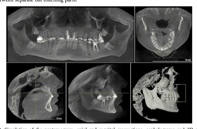
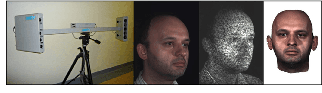
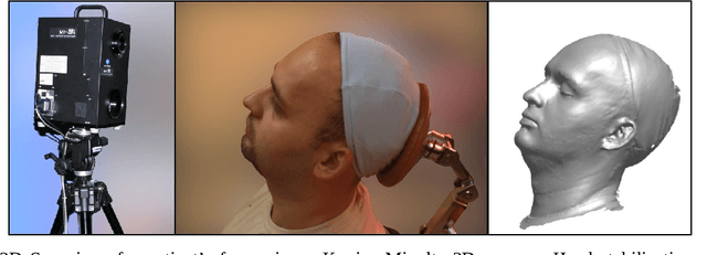
Apr 17, 2025Contemporary digital technology has a pivotal role in the design of customized medical appliances, including occlusal splints used in the treatment of stomatognathic system dysfunctions. We present an approach to computer-aided design and precision assessment of positioning occlusal splints, bridging clinical concepts with current digital dental practice. In our model, a 3D splint is generated based on a transformation matrix that represents the therapeutic change in mandibular position, defined by a specialist using a virtual patient model reconstructed from intraoral scans, CBCT, 3D facial scans and plaster model digitisation. The paper introduces a novel method for generating splints that accurately reproduce occlusal conditions in the therapeutic position, including a mechanism for resolving surface conflicts through virtual embossing. We demonstrate how transformation matrices can be acquired through clinical tools and intraoral devices, and evaluate the accuracy of the designed and printed splints using profile and surface deviation analysis. The proposed method enables reproducible, patient-specific splint fabrication and opens new possibilities in diagnostics, multimodal image registration and quantification of occlusal discrepancies.
Transformation trees -- documentation of multimodal image registration
Jan 31, 2025The paper presents proposals for the application of a tree structure to the documentation of a set of transformations obtained as a result of various registrations of multimodal images obtained in coordinate systems associated with acquisition devices and being registered in one patient-specific coordinate system. A special file format .dpw (digital patient workspace) is introduced. Examples of different registrations yielded from orthodontic analysis and showing main aspects of the usage of tree structure are illustrated in dpVision software.
The dynamics of the stomatognathic system from 4D multimodal data
Nov 20, 2019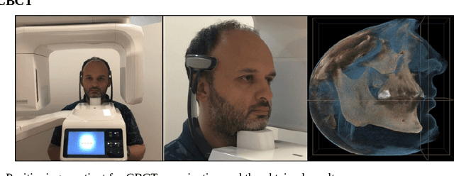
The purpose of this chapter is to discuss methods of acquisition, visualization and analysis of the dynamics of a complex biomedical system, illustrated by the human stomatognathic system. The stomatognathic system consists of the teeth and the skull bones with the maxilla and the mandible. Its dynamics can be described by the change of mutual position of the lower/mandibular part versus the upper/maxillary one due to the physiological motion of opening, chewing and swallowing. In order to analyse the dynamics of the stomatognathic system its morphology and motion has to be digitized, which is done using static and dynamic multimodal imagery like CBCT and 3D scans data and temporal measurements of motion. The integration of multimodal data incorporates different direct and indirect methods of registration - aligning of all the data in the same coordinate system. The integrated sets of data form 4D multimodal data which can be further visualized, modeled, and subjected to multivariate time series analysis. Example results are shown. Although there is no direct method of imaging the TMJ motion, the integration of multimodal data forms an adequate tool. As medical imaging becomes ever more diverse and ever more accessible, organizing the imagery and measurements into unified, comprehensive records can deliver to the doctor the most information in the most accessible form, creating a new quality in data simulation, analysis and interpretation.