 Add to Chrome
 Add to Firefox
 Add to Edge
Transformation-driven generation of comparable projection images from multimodal anatomical scenes
Jun 15, 2026This work addresses the computational problem of generating reproducible projection-space observations from heterogeneous anatomical scenes whose components may undergo independent spatial transformations. We propose a transformation-driven framework for synthetic projection imaging from multimodal anatomical data and demonstrate it on mandibular-motion scenarios. In contrast to conventional Digitally Reconstructed Radiograph (DRR) approaches primarily designed for registration, projection realism, or rendering efficiency, the proposed formulation treats projection imaging as an observation process operating on an explicitly represented anatomical scene. Independently transformable volumetric and surface-based anatomical objects are embedded within a shared scene representation and propagated directly into projection space through explicit transformations. Projection geometry, acquisition modelling, material interpretation, and image presentation remain explicitly separated, enabling controlled exploration of methodological assumptions while preserving reproducibility and direct comparability between generated projections. Particular emphasis is placed on transformation-driven anatomical scenarios relevant to craniofacial analysis, including mandibular motion and therapeutic repositioning. Using a shared anatomical reference scene composed of CT/CBCT volumes, segmented structures, surface models, and auxiliary anatomical or therapeutic objects, the framework enables generation of directly comparable VirtualRTG projections from multiple anatomical configurations while preserving identical imaging assumptions. Rather than aiming at fully physically faithful radiographic simulation, the proposed approach provides a controllable and reproducible methodological environment for studying anatomy--projection relationships, motion observability, and transformation-aware imaging workflows.
Assessment of the quantitative impact of occlusal positioning splints on temporomandibular joint conditions
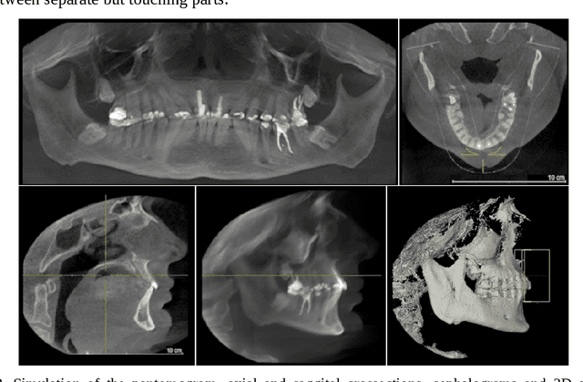
Apr 28, 2026A computational method for quantitative analysis of temporomandibular joint (TMJ) configuration using occlusal positioning splints is proposed and demonstrated. The method models a positioning splint as a physical realization of a predefined rigid transformation of the mandible, derived from multimodal data, including CBCT, facial motion acquisition, and dental scans integrated within a common coordinate system. Splints corresponding to selected mandibular positions are designed and fabricated, and their positioning accuracy is evaluated using repeated scans of plaster models. Discrepancies are represented as error transformations and analyzed statistically in the space of rigid motions. The estimated transformations are propagated to segmented TMJ structures, enabling simulation-based evaluation of joint space changes. Transformation-based error analysis and surface distance metrics are used to quantify differences between planned and achieved configurations. The method enables indirect assessment of TMJ configuration using a single anatomical model and transformation data, reducing the need for repeated imaging across multiple mandibular positions. This study is intended as a methodological demonstration, supported by a clear step-by-step graphical presentation, and does not aim to provide clinical validation.
Computer-Aided Design of Personalized Occlusal Positioning Splints Using Multimodal 3D Data
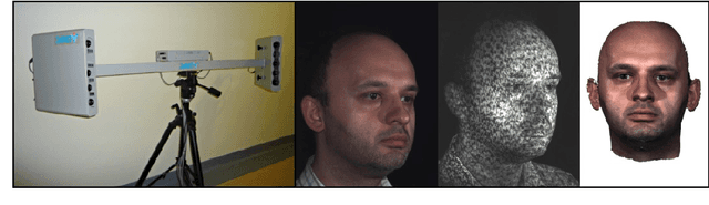
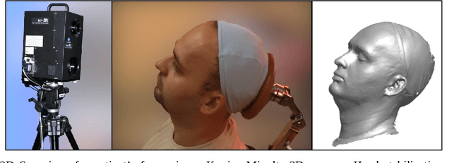
Apr 17, 2025Contemporary digital technology has a pivotal role in the design of customized medical appliances, including occlusal splints used in the treatment of stomatognathic system dysfunctions. We present an approach to computer-aided design and precision assessment of positioning occlusal splints, bridging clinical concepts with current digital dental practice. In our model, a 3D splint is generated based on a transformation matrix that represents the therapeutic change in mandibular position, defined by a specialist using a virtual patient model reconstructed from intraoral scans, CBCT, 3D facial scans and plaster model digitisation. The paper introduces a novel method for generating splints that accurately reproduce occlusal conditions in the therapeutic position, including a mechanism for resolving surface conflicts through virtual embossing. We demonstrate how transformation matrices can be acquired through clinical tools and intraoral devices, and evaluate the accuracy of the designed and printed splints using profile and surface deviation analysis. The proposed method enables reproducible, patient-specific splint fabrication and opens new possibilities in diagnostics, multimodal image registration and quantification of occlusal discrepancies.
Visualisation of a multidimensional point cloud as a 3D swarm of avatars
Apr 09, 2025The article presents an innovative approach to the visualisation of multidimensional data, using icons inspired by Chernoff faces. The approach merges classical projection techniques with the assignment of particular data dimensions to mimic features, capitalizing on the natural ability of the human brain to interpret facial expressions. The technique is implemented as a plugin to the dpVision open-source image handling platform. The plugin allows the data to be interactively explored in the form of a swarm of "totems" whose position in hyperspace as well as facial features represent various aspects of the data. Sample visualisations, based on synthetic test data as well as the vinhoverde 15-dimensional database on Portuguese wines, confirm the usefulness of our approach to the analysis of complex data structures.
Transformation trees -- documentation of multimodal image registration
Jan 31, 2025The paper presents proposals for the application of a tree structure to the documentation of a set of transformations obtained as a result of various registrations of multimodal images obtained in coordinate systems associated with acquisition devices and being registered in one patient-specific coordinate system. A special file format .dpw (digital patient workspace) is introduced. Examples of different registrations yielded from orthodontic analysis and showing main aspects of the usage of tree structure are illustrated in dpVision software.
The dynamics of the stomatognathic system from 4D multimodal data
Nov 20, 2019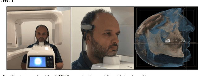
The purpose of this chapter is to discuss methods of acquisition, visualization and analysis of the dynamics of a complex biomedical system, illustrated by the human stomatognathic system. The stomatognathic system consists of the teeth and the skull bones with the maxilla and the mandible. Its dynamics can be described by the change of mutual position of the lower/mandibular part versus the upper/maxillary one due to the physiological motion of opening, chewing and swallowing. In order to analyse the dynamics of the stomatognathic system its morphology and motion has to be digitized, which is done using static and dynamic multimodal imagery like CBCT and 3D scans data and temporal measurements of motion. The integration of multimodal data incorporates different direct and indirect methods of registration - aligning of all the data in the same coordinate system. The integrated sets of data form 4D multimodal data which can be further visualized, modeled, and subjected to multivariate time series analysis. Example results are shown. Although there is no direct method of imaging the TMJ motion, the integration of multimodal data forms an adequate tool. As medical imaging becomes ever more diverse and ever more accessible, organizing the imagery and measurements into unified, comprehensive records can deliver to the doctor the most information in the most accessible form, creating a new quality in data simulation, analysis and interpretation.