 Add to Chrome
 Add to Firefox
 Add to Edge
Geometric Deep Learning for Automated Landmarking of Maxillary Arches on 3D Oral Scans from Newborns with Cleft Lip and Palate
Jan 27, 2025


Rapid advances in 3D model scanning have enabled the mass digitization of dental clay models. However, most clinicians and researchers continue to use manual morphometric analysis methods on these models such as landmarking. This is a significant step in treatment planning for craniomaxillofacial conditions. We aimed to develop and test a geometric deep learning model that would accurately and reliably label landmarks on a complicated and specialized patient population -- infants, as accurately as a human specialist without a large amount of training data. Our developed pipeline demonstrated an accuracy of 94.44% with an absolute mean error of 1.676 +/- 0.959 mm on a set of 100 models acquired from newborn babies with cleft lip and palate. Our proposed pipeline has the potential to serve as a fast, accurate, and reliable quantifier of maxillary arch morphometric features, as well as an integral step towards a future fully automated dental treatment pipeline.
The Brain Tumor Segmentation Challenge 2023: Glioma Segmentation in Sub-Saharan Africa Patient Population
May 30, 2023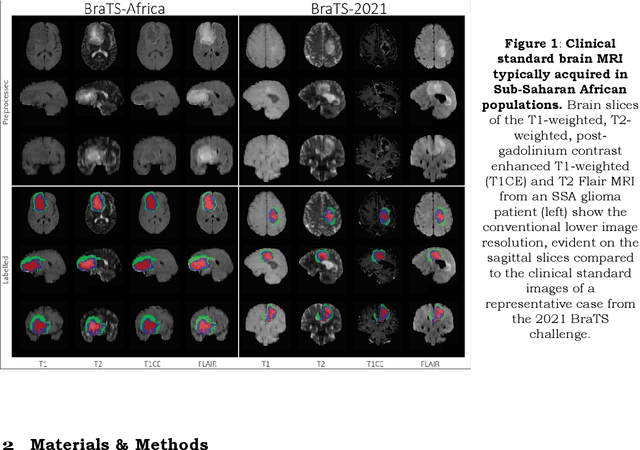
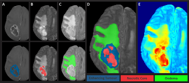
Gliomas are the most common type of primary brain tumors. Although gliomas are relatively rare, they are among the deadliest types of cancer, with a survival rate of less than 2 years after diagnosis. Gliomas are challenging to diagnose, hard to treat and inherently resistant to conventional therapy. Years of extensive research to improve diagnosis and treatment of gliomas have decreased mortality rates across the Global North, while chances of survival among individuals in low- and middle-income countries (LMICs) remain unchanged and are significantly worse in Sub-Saharan Africa (SSA) populations. Long-term survival with glioma is associated with the identification of appropriate pathological features on brain MRI and confirmation by histopathology. Since 2012, the Brain Tumor Segmentation (BraTS) Challenge have evaluated state-of-the-art machine learning methods to detect, characterize, and classify gliomas. However, it is unclear if the state-of-the-art methods can be widely implemented in SSA given the extensive use of lower-quality MRI technology, which produces poor image contrast and resolution and more importantly, the propensity for late presentation of disease at advanced stages as well as the unique characteristics of gliomas in SSA (i.e., suspected higher rates of gliomatosis cerebri). Thus, the BraTS-Africa Challenge provides a unique opportunity to include brain MRI glioma cases from SSA in global efforts through the BraTS Challenge to develop and evaluate computer-aided-diagnostic (CAD) methods for the detection and characterization of glioma in resource-limited settings, where the potential for CAD tools to transform healthcare are more likely.