 Add to Chrome
 Add to Firefox
 Add to Edge
Coronary Artery Segmentation and Vessel-Type Classification in X-Ray Angiography
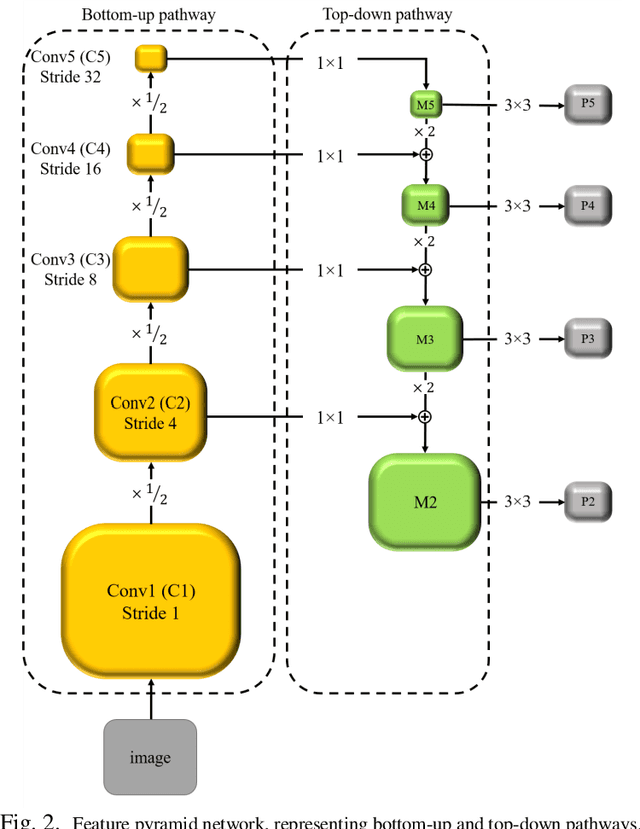
Jan 24, 2026X-ray coronary angiography (XCA) is the clinical reference standard for assessing coronary artery disease, yet quantitative analysis is limited by the difficulty of robust vessel segmentation in routine data. Low contrast, motion, foreshortening, overlap, and catheter confounding degrade segmentation and contribute to domain shift across centers. Reliable segmentation, together with vessel-type labeling, enables vessel-specific coronary analytics and downstream measurements that depend on anatomical localization. From 670 cine sequences (407 subjects), we select a best frame near peak opacification using a low-intensity histogram criterion and apply joint super-resolution and enhancement. We benchmark classical Meijering, Frangi, and Sato vesselness filters under per-image oracle tuning, a single global mean setting, and per-image parameter prediction via Support Vector Regression (SVR). Neural baselines include U-Net, FPN, and a Swin Transformer, trained with coronary-only and merged coronary+catheter supervision. A second stage assigns vessel identity (LAD, LCX, RCA). External evaluation uses the public DCA1 cohort. SVR per-image tuning improves Dice over global means for all classical filters (e.g., Frangi: 0.759 vs. 0.741). Among deep models, FPN attains 0.914+/-0.007 Dice (coronary-only), and merged coronary+catheter labels further improve to 0.931+/-0.006. On DCA1 as a strict external test, Dice drops to 0.798 (coronary-only) and 0.814 (merged), while light in-domain fine-tuning recovers to 0.881+/-0.014 and 0.882+/-0.015. Vessel-type labeling achieves 98.5% accuracy (Dice 0.844) for RCA, 95.4% (0.786) for LAD, and 96.2% (0.794) for LCX. Learned per-image tuning strengthens classical pipelines, while high-resolution FPN models and merged-label supervision improve stability and external transfer with modest adaptation.
MFP-Unet: A Novel Deep Learning Based Approach for Left Ventricle Segmentation in Echocardiography
Jun 25, 2019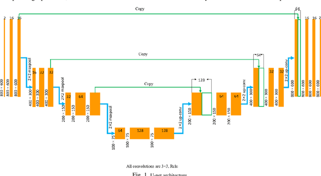
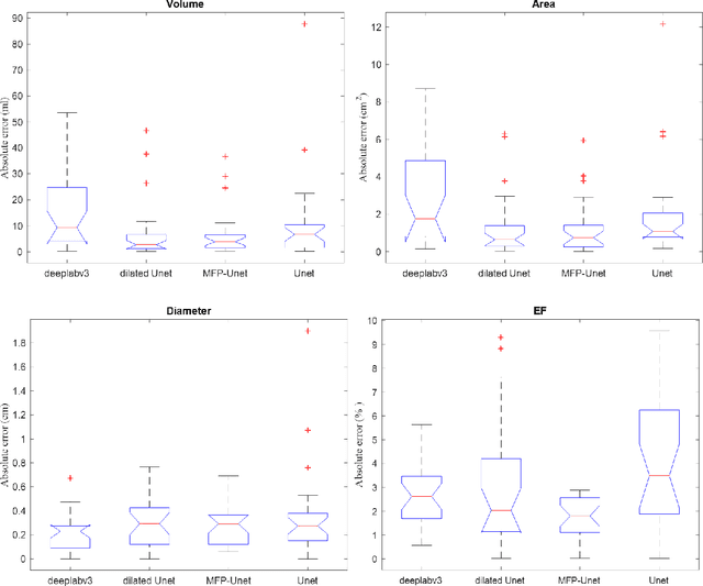
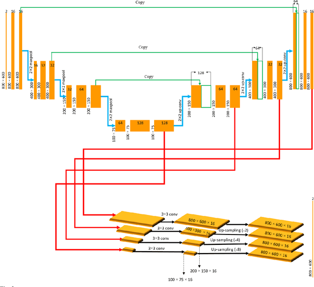
Segmentation of the Left ventricle (LV) is a crucial step for quantitative measurements such as area, volume, and ejection fraction. However, the automatic LV segmentation in 2D echocardiographic images is a challenging task due to ill-defined borders, and operator dependence issues (insufficient reproducibility). U-net, which is a well-known architecture in medical image segmentation, addressed this problem through an encoder-decoder path. Despite outstanding overall performance, U-net ignores the contribution of all semantic strengths in the segmentation procedure. In the present study, we have proposed a novel architecture to tackle this drawback. Feature maps in all levels of the decoder path of U-net are concatenated, their depths are equalized, and up-sampled to a fixed dimension. This stack of feature maps would be the input of the semantic segmentation layer. The proposed network yielded state-of-the-art results when comparing with results from U-net, dilated U-net, and deeplabv3, using the same dataset. An average Dice Metric (DM) of 0.945, Hausdorff Distance (HD) of 1.62, Jaccard Coefficient (JC) of 0.97, and Mean Absolute Distance (MAD) of 1.32 are achieved. The correlation graph, bland-altman analysis, and box plot showed a great agreement between automatic and manually calculated volume, area, and length.