Klaus Engelke
A New 3D Segmentation Technique for QCT Scans of the Lumbar Spine to Determine BMD and Vertebral Geometry
May 19, 2017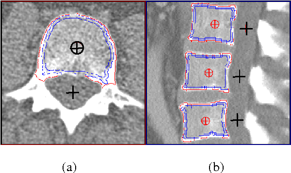
Abstract:Quantitative computed tomography (QCT) is a standard method to determine bone mineral density (BMD) in the spine. Traditionally single 8 - 10 mm thick slices have been analyzed only. Current spiral CT scanners provide true 3D acquisition schemes allowing for a more differential BMD analysis and an assessment of geometric parameters, which may improve fracture prediction. We developed a novel 3D segmentation approach that combines deformable balloons, multi seeded volume growing, and dedicated morphological operations to extract the vertebral bodies. An anatomy-oriented coordinate system attached automatically to each vertebra is used to define volumes of interest. We analyzed intra-operator precision of the segmentation procedure using abdominal scans from 10 patients (60 mAs, 120 kV, slice thickness 1mm, B40s, Siemens Sensation 16). Our new segmentation method shows excellent precision errors in the order of < 1 % for BMD and < 2 % for volume.
A New 3D Method to Segment the Lumbar Vertebral Bodies and to Determine Bone Mineral Density and Geometry
May 19, 2017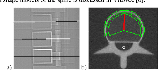
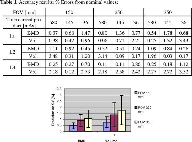


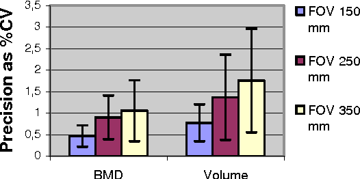
Abstract:In this paper we present a new 3D segmentation approach for the vertebrae of the lower thoracic and the lumbar spine in spiral computed tomography datasets. We implemented a multi-step procedure. Its main components are deformable models, volume growing, and morphological operations. The performance analysis that included an evaluation of accuracy using the European Spine Phantom, and of intra-operator precision using clinical CT datasets from 10 patients highlight the potential for clinical use. The intra-operator precision of the segmentation procedure was better than 1% for Bone Mineral Density (BMD) and better than 1.8% for volume. The long-term goal of this work is to enable better fracture prediction and improved patient monitoring in the field of osteoporosis. A true 3D segmentation also enables an accurate measurement of geometrical parameters that can augment the classical measurement of BMD.
A New 3D Segmentation Methodology for Lumbar Vertebral Bodies for the Measurement of BMD and Geometry
May 19, 2017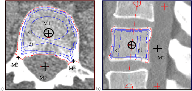

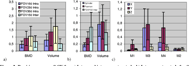
Abstract:In this paper a new technique is presented that extracts the geometry of lumbar vertebral bodies from spiral CT scans. Our new multi-step segmentation approach yields highly accurate and precise measurement of the bone mineral density (BMD) in different volumes of interest which are defined relative to a local anatomical coordinate systems. The approach also enables the analysis of the geometry of the relevant vertebrae. Intra- and inter operator precision for segmentation, BMD measurement and position of the coordinate system are below 1.5% in patient data, accuracy errors are below 1.5% for BMD and below 4% for volume in phantom data. The long-term goal of the approach is to improve fracture prediction in osteoporosis.
 Add to Chrome
 Add to Firefox
 Add to Edge