A New 3D Segmentation Methodology for Lumbar Vertebral Bodies for the Measurement of BMD and Geometry
Paper and Code
May 19, 2017


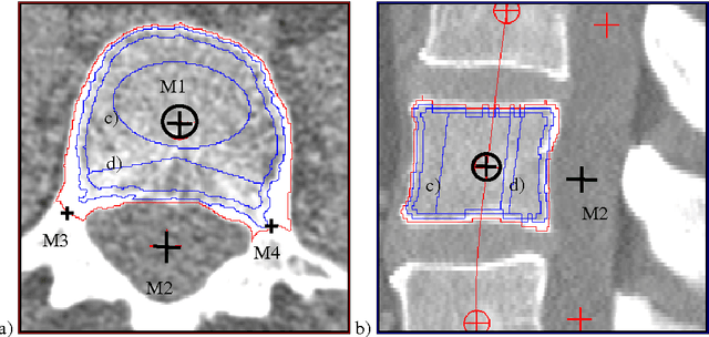
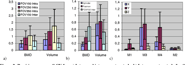
In this paper a new technique is presented that extracts the geometry of lumbar vertebral bodies from spiral CT scans. Our new multi-step segmentation approach yields highly accurate and precise measurement of the bone mineral density (BMD) in different volumes of interest which are defined relative to a local anatomical coordinate systems. The approach also enables the analysis of the geometry of the relevant vertebrae. Intra- and inter operator precision for segmentation, BMD measurement and position of the coordinate system are below 1.5% in patient data, accuracy errors are below 1.5% for BMD and below 4% for volume in phantom data. The long-term goal of the approach is to improve fracture prediction in osteoporosis.
* 4 pages,2 figures, MIUA05 conference
 Add to Chrome
 Add to Firefox
 Add to Edge