 Add to Chrome
 Add to Firefox
 Add to Edge
Expert Consensus on Criteria for the Automated Assessment of Laparoscopic Camera Navigation
Jun 22, 2026
Background: Laparoscopic camera navigation (LCN) is a critical skill, yet its current assessment typically relies on manual rating systems which are time-consuming and difficult to scale. Automated feedback could significantly enhance surgical training by providing immediate, standardized metrics. This study aims to define, clinically evaluate the relevance, and establish the technical readiness of a set of approaches for LCN assessment. Methods: We developed a detailed taxonomy of 14 key aspects of camera navigation, categorized into Framing & Composition, Visibility & Clarity, Orientation & Stability, Motion & Dynamics, and Safety & Awareness. For each aspect, we assessed the technological readiness of automated measurement based on the current state of the art (SoTA) in computer vision (CV). To establish clinical relevance, we designed a survey for practicing laparoscopic surgeons to rate the importance of each aspect on a 5-point Likert scale and to select the five most critical skills. Results: 23 surgeons participated in the survey. Foundational aspects like Field of View, Focus and Centering were rated as most important by surgeons. We present a "Clinical Importance vs. CV Technological Readiness" matrix, identifying high-priority targets for development--aspects that are both clinically crucial and technologically ready to measure. Conclusion: This work establishes a foundational framework for quantifying LCN skills. By aligning surgeon priorities with CV capabilities, we provide a clear roadmap for automatic skill assessment. This foundation enables the development of AI-driven assistance tools that can accelerate the learning curve for surgical assistants and potentially improve surgical safety and efficiency.

Comparison of Representation Learning Techniques for Tracking in time resolved 3D Ultrasound
Jan 10, 2022
3D ultrasound (3DUS) becomes more interesting for target tracking in radiation therapy due to its capability to provide volumetric images in real-time without using ionizing radiation. It is potentially usable for tracking without using fiducials. For this, a method for learning meaningful representations would be useful to recognize anatomical structures in different time frames in representation space (r-space). In this study, 3DUS patches are reduced into a 128-dimensional r-space using conventional autoencoder, variational autoencoder and sliced-wasserstein autoencoder. In the r-space, the capability of separating different ultrasound patches as well as recognizing similar patches is investigated and compared based on a dataset of liver images. Two metrics to evaluate the tracking capability in the r-space are proposed. It is shown that ultrasound patches with different anatomical structures can be distinguished and sets of similar patches can be clustered in r-space. The results indicate that the investigated autoencoders have different levels of usability for target tracking in 3DUS.
Robotized Ultrasound Imaging of the Peripheral Arteries -- a Phantom Study
Jul 13, 2020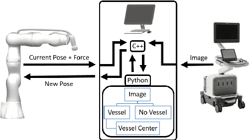
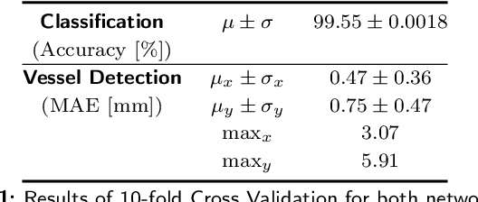
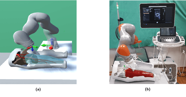
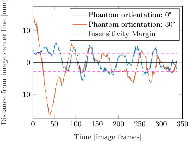
The first choice in diagnostic imaging for patients suffering from peripheral arterial disease is 2D ultrasound (US). However, for a proper imaging process, a skilled and experienced sonographer is required. Additionally, it is a highly user-dependent operation. A robotized US system that autonomously scans the peripheral arteries has the potential to overcome these limitations. In this work, we extend a previously proposed system by a hierarchical image analysis pipeline based on convolutional neural networks in order to control the robot. The system was evaluated by checking its feasibility to keep the vessel lumen of a leg phantom within the US image while scanning along the artery. In 100 % of the images acquired during the scan process the whole vessel lumen was visible. While defining an insensitivity margin of 2.74 mm, the mean absolute distance between vessel center and the horizontal image center line was 2.47 mm and 3.90 mm for an easy and complex scenario, respectively. In conclusion, this system presents the basis for fully automatized peripheral artery imaging in humans using a radiation-free approach.