Francisco Javier Díaz-Pernas
A Deep Learning Approach for Brain Tumor Classification and Segmentation Using a Multiscale Convolutional Neural Network
Feb 04, 2024

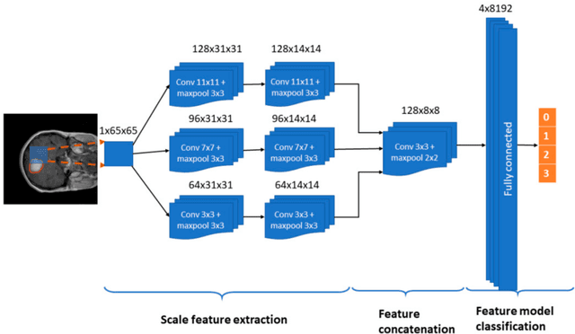

Abstract:In this paper, we present a fully automatic brain tumor segmentation and classification model using a Deep Convolutional Neural Network that includes a multiscale approach. One of the differences of our proposal with respect to previous works is that input images are processed in three spatial scales along different processing pathways. This mechanism is inspired in the inherent operation of the Human Visual System. The proposed neural model can analyze MRI images containing three types of tumors: meningioma, glioma, and pituitary tumor, over sagittal, coronal, and axial views and does not need preprocessing of input images to remove skull or vertebral column parts in advance. The performance of our method on a publicly available MRI image dataset of 3064 slices from 233 patients is compared with previously classical machine learning and deep learning published methods. In the comparison, our method remarkably obtained a tumor classification accuracy of 0.973, higher than the other approaches using the same database.
* 14 pages
A comparative study on wearables and single-camera video for upper-limb out-of-thelab activity recognition with different deep learning architectures
Feb 04, 2024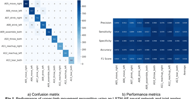
Abstract:The use of a wide range of computer vision solutions, and more recently high-end Inertial Measurement Units (IMU) have become increasingly popular for assessing human physical activity in clinical and research settings. Nevertheless, to increase the feasibility of patient tracking in out-of-the-lab settings, it is necessary to use a reduced number of devices for movement acquisition. Promising solutions in this context are IMU-based wearables and single camera systems. Additionally, the development of machine learning systems able to recognize and digest clinically relevant data in-the-wild is needed, and therefore determining the ideal input to those is crucial.
A Physiological Sensor-Based Android Application Synchronized with a Driving Simulator for Driver Monitoring
Feb 04, 2024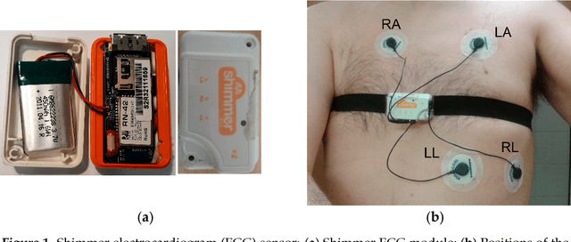
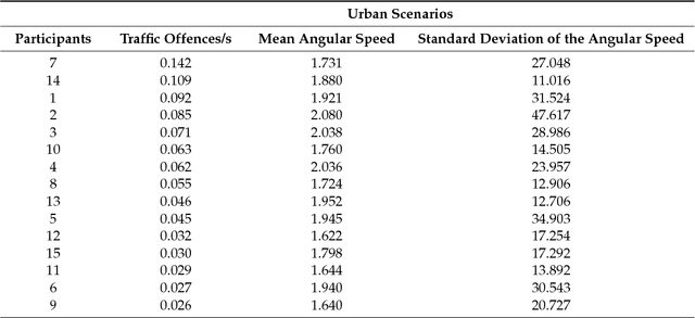
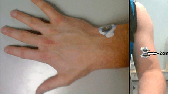
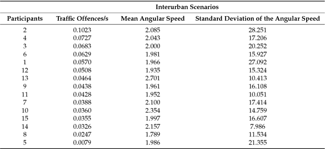
Abstract:In this paper, we present an Android application to control and monitor the physiological sensors from the Shimmer platform and its synchronized working with a driving simulator. The Android app can monitor drivers and their parameters can be used to analyze the relation between their physiological states and driving performance. The app can configure, select, receive, process, represent graphically, and store the signals from electrocardiogram (ECG), electromyogram (EMG) and galvanic skin response (GSR) modules and accelerometers, a magnetometer and a gyroscope. The Android app is synchronized in two steps with a driving simulator that we previously developed using the Unity game engine to analyze driving security and efficiency. The Android app was tested with different sensors working simultaneously at various sampling rates and in different Android devices. We also tested the synchronized working of the driving simulator and the Android app with 25 people and analyzed the relation between data from the ECG, EMG, GSR, and gyroscope sensors and from the simulator. Among others, some significant correlations between a gyroscope-based feature calculated by the Android app and vehicle data and particular traffic offences were found. The Android app can be applied with minor adaptations to other different users such as patients with chronic diseases or athletes.
* 28 pages
 Add to Chrome
 Add to Firefox
 Add to Edge