Fabrizio Zanconati
Hybrid quantum image classification and federated learning for hepatic steatosis diagnosis
Nov 04, 2023


Abstract:With the maturity achieved by deep learning techniques, intelligent systems that can assist physicians in the daily interpretation of clinical images can play a very important role. In addition, quantum techniques applied to deep learning can enhance this performance, and federated learning techniques can realize privacy-friendly collaborative learning among different participants, solving privacy issues due to the use of sensitive data and reducing the number of data to be collected for each individual participant. We present in this study a hybrid quantum neural network that can be used to quantify non-alcoholic liver steatosis and could be useful in the diagnostic process to determine a liver's suitability for transplantation; at the same time, we propose a federated learning approach based on a classical deep learning solution to solve the same problem, but using a reduced data set in each part. The liver steatosis image classification accuracy of the hybrid quantum neural network, the hybrid quantum ResNet model, consisted of 5 qubits and more than 100 variational gates, reaches 97%, which is 1.8% higher than its classical counterpart, ResNet. Crucially, that even with a reduced dataset, our hybrid approach consistently outperformed its classical counterpart, indicating superior generalization and less potential for overfitting in medical applications. In addition, a federated approach with multiple clients, up to 32, despite the lower accuracy, but still higher than 90%, would allow using, for each participant, a very small dataset, i.e., up to one-thirtieth. Our work, based over real-word clinical data can be regarded as a scalable and collaborative starting point, could thus fulfill the need for an effective and reliable computer-assisted system that facilitates the daily diagnostic work of the clinical pathologist.
Tomographic phase and attenuation extraction for a sample composed of unknown materials using X-ray propagation-based phase-contrast imaging
Oct 14, 2021


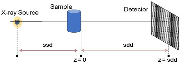
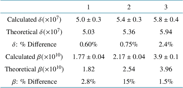
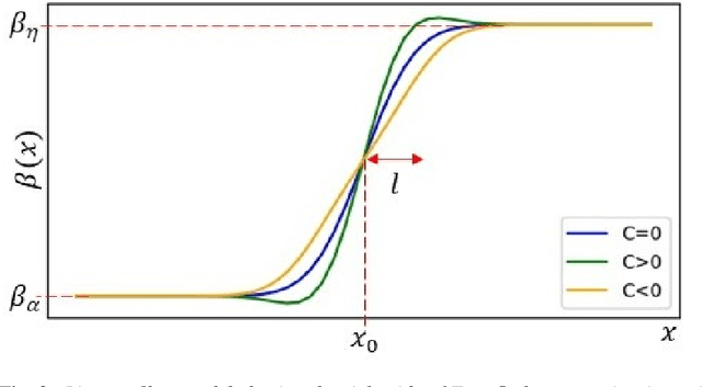
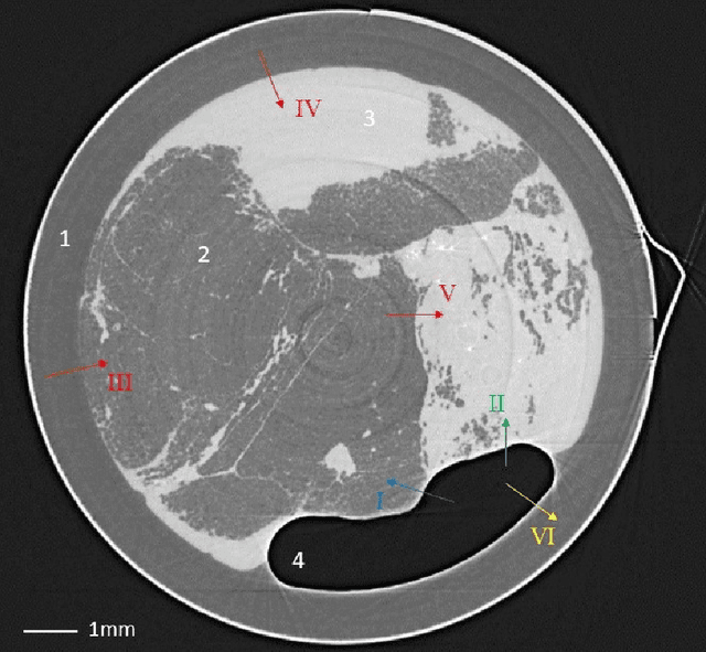
Abstract:Propagation-based phase-contrast X-ray imaging (PB-PCXI) generates image contrast by utilizing sample-imposed phase-shifts. This has proven useful when imaging weakly-attenuating samples, as conventional attenuation-based imaging does not always provide adequate contrast. We present a PB-PCXI algorithm capable of extracting the X-ray attenuation, $\beta$, and refraction, $\delta$, components of the complex refractive index of distinct materials within an unknown sample. The method involves curve-fitting an error-function-based model to a phase-retrieved interface in a PB-PCXI tomographic reconstruction, which is obtained when Paganin-type phase-retrieval is applied with incorrect values of $\delta$ and $\beta$. The fit parameters can then be used to calculate true $\delta$ and $\beta$ values for composite materials. This approach requires no a priori sample information, making it broadly applicable. Our PB-PCXI reconstruction is single distance, requiring only one exposure per tomographic angle, which is important for radiosensitive samples. We apply this approach to a breast-tissue sample, recovering the refraction component, $\delta$, with 0.6 - 2.4\% accuracy compared to theoretical values.
 Add to Chrome
 Add to Firefox
 Add to Edge