Emad-ul-Haq Qazi
Automatic Emotion Recognition (AER) System based on Two-Level Ensemble of Lightweight Deep CNN Models
Apr 30, 2019


Abstract:Emotions play a crucial role in human interaction, health care and security investigations and monitoring. Automatic emotion recognition (AER) using electroencephalogram (EEG) signals is an effective method for decoding the real emotions, which are independent of body gestures, but it is a challenging problem. Several automatic emotion recognition systems have been proposed, which are based on traditional hand-engineered approaches and their performances are very poor. Motivated by the outstanding performance of deep learning (DL) in many recognition tasks, we introduce an AER system (Deep-AER) based on EEG brain signals using DL. A DL model involves a large number of learnable parameters, and its training needs a large dataset of EEG signals, which is difficult to acquire for AER problem. To overcome this problem, we proposed a lightweight pyramidal one-dimensional convolutional neural network (LP-1D-CNN) model, which involves a small number of learnable parameters. Using LP-1D-CNN, we build a two level ensemble model. In the first level of the ensemble, each channel is scanned incrementally by LP-1D-CNN to generate predictions, which are fused using majority vote. The second level of the ensemble combines the predictions of all channels of an EEG signal using majority vote for detecting the emotion state. We validated the effectiveness and robustness of Deep-AER using DEAP, a benchmark dataset for emotion recognition research. The results indicate that FRONT plays dominant role in AER and over this region, Deep-AER achieved the accuracies of 98.43% and 97.65% for two AER problems, i.e., high valence vs low valence (HV vs LV) and high arousal vs low arousal (HA vs LA), respectively. The comparison reveals that Deep-AER outperforms the state-of-the-art systems with large margin. The Deep-AER system will be helpful in monitoring for health care and security investigations.
An Efficient Intelligent System for the Classification of Electroencephalography (EEG) Brain Signals using Nuclear Features for Human Cognitive Tasks
Apr 30, 2019


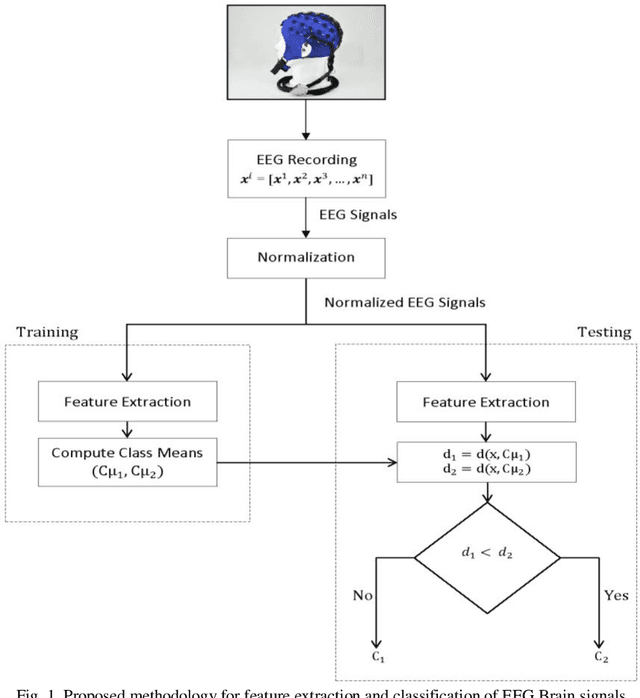
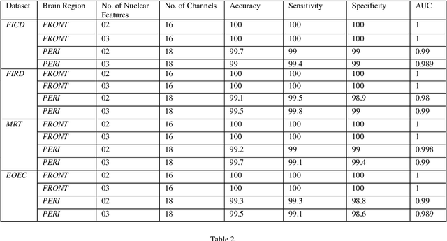
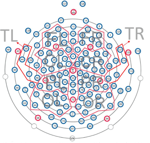
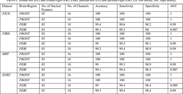
Abstract:Representation and classification of Electroencephalography (EEG) brain signals are critical processes for their analysis in cognitive tasks. Particularly, extraction of discriminative features from raw EEG signals, without any pre-processing, is a challenging task. Motivated by nuclear norm, we observed that there is a significant difference between the variances of EEG signals captured from the same brain region when a subject performs different tasks. This observation lead us to use singular value decomposition for computing dominant variances of EEG signals captured from a certain brain region while performing a certain task and use them as features (nuclear features). A simple and efficient class means based minimum distance classifier (CMMDC) is enough to predict brain states. This approach results in the feature space of significantly small dimension and gives equally good classification results on clean as well as raw data. We validated the effectiveness and robustness of the technique using four datasets of different tasks: fluid intelligence clean data (FICD), fluid intelligence raw data (FIRD), memory recall task (MRT), and eyes open / eyes closed task (EOEC). For each task, we analyzed EEG signals over six (06) different brain regions with 8, 16, 20, 18, 18 and 100 electrodes. The nuclear features from frontal brain region gave the 100% prediction accuracy. The discriminant analysis of the nuclear features has been conducted using intra-class and inter-class variations. Comparisons with the state-of-the-art techniques showed the superiority of the proposed system.
Eigen Values Features for the Classification of Brain Signals corresponding to 2D and 3D Educational Contents
Apr 30, 2019


Abstract:In this paper, we have proposed a brain signal classification method, which uses eigenvalues of the covariance matrix as features to classify images (topomaps) created from the brain signals. The signals are recorded during the answering of 2D and 3D questions. The system is used to classify the correct and incorrect answers for both 2D and 3D questions. Using the classification technique, the impacts of 2D and 3D multimedia educational contents on learning, memory retention and recall will be compared. The subjects learn similar 2D and 3D educational contents. Afterwards, subjects are asked 20 multiple-choice questions (MCQs) associated with the contents after thirty minutes (Short-Term Memory) and two months (Long-Term Memory). Eigenvalues features extracted from topomaps images are given to K-Nearest Neighbor (KNN) and Support Vector Machine (SVM) classifiers, in order to identify the states of the brain related to incorrect and correct answers. Excellent accuracies obtained by both classifiers and by applying statistical analysis on the results, no significant difference is indicated between 2D and 3D multimedia educational contents on learning, memory retention and recall in both STM and LTM.
An Automated System for Epilepsy Detection using EEG Brain Signals based on Deep Learning Approach
Jan 16, 2018


Abstract:Epilepsy is a neurological disorder and for its detection, encephalography (EEG) is a commonly used clinical approach. Manual inspection of EEG brain signals is a time-consuming and laborious process, which puts heavy burden on neurologists and affects their performance. Several automatic techniques have been proposed using traditional approaches to assist neurologists in detecting binary epilepsy scenarios e.g. seizure vs. non-seizure or normal vs. ictal. These methods do not perform well when classifying ternary case e.g. ictal vs. normal vs. inter-ictal; the maximum accuracy for this case by the state-of-the-art-methods is 97+-1%. To overcome this problem, we propose a system based on deep learning, which is an ensemble of pyramidal one-dimensional convolutional neural network (P-1D-CNN) models. In a CNN model, the bottleneck is the large number of learnable parameters. P-1D-CNN works on the concept of refinement approach and it results in 60% fewer parameters compared to traditional CNN models. Further to overcome the limitations of small amount of data, we proposed augmentation schemes for learning P-1D-CNN model. In almost all the cases concerning epilepsy detection, the proposed system gives an accuracy of 99.1+-0.9% on the University of Bonn dataset.
 Add to Chrome
 Add to Firefox
 Add to Edge