Claas Strodthoff
Classification of Electrical Impedance Tomography Data Using Machine Learning
Aug 02, 2021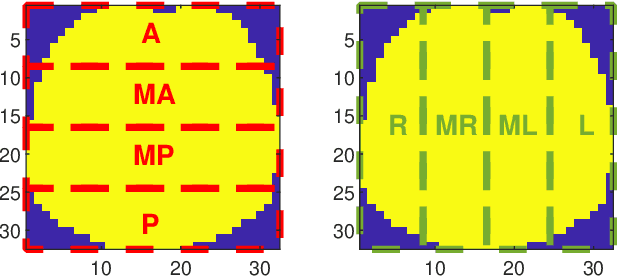
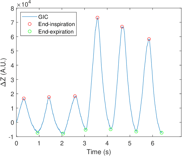
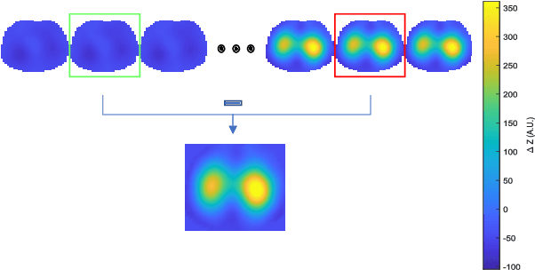
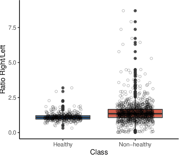
Abstract:Patients suffering from pulmonary diseases typically exhibit pathological lung ventilation in terms of homogeneity. Electrical Impedance Tomography (EIT) is a non-invasive imaging method that allows to analyze and quantify the distribution of ventilation in the lungs. In this article, we present a new approach to promote the use of EIT data and the implementation of new clinical applications for differential diagnosis, with the development of several machine learning models to discriminate between EIT data from healthy and non-healthy subjects. EIT data from 16 subjects were acquired: 5 healthy and 11 non-healthy subjects (with multiple pulmonary conditions). Preliminary results have shown accuracy percentages of 66\% in challenging evaluation scenarios. The results suggest that the pairing of EIT feature engineering methods with machine learning methods could be further explored and applied in the diagnostic and monitoring of patients suffering from lung diseases. Also, we introduce the use of a new feature in the context of EIT data analysis (Impedance Curve Correlation).
Inferring respiratory and circulatory parameters from electrical impedance tomography with deep recurrent models
Oct 19, 2020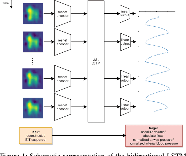
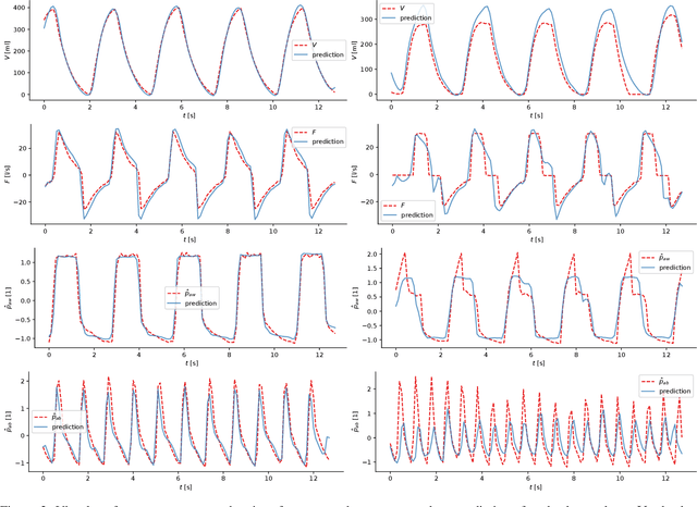
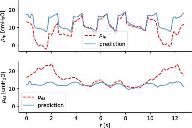

Abstract:Electrical impedance tomography (EIT) is a noninvasive imaging modality that allows a continuous assessment of changes in regional bioimpedance of different organs. One of its most common biomedical applications is monitoring regional ventilation distribution in critically ill patients treated in intensive care units. In this work, we put forward a proof-of-principle study that demonstrates how one can reconstruct synchronously measured respiratory or circulatory parameters from the EIT image sequence using a deep learning model trained in an end-to-end fashion. We demonstrate that one can accurately infer absolute volume, absolute flow, normalized airway pressure and within certain limitations even the normalized arterial blood pressure from the EIT signal alone, in a way that generalizes to unseen patients without prior calibration. As an outlook with direct clinical relevance, we furthermore demonstrate the feasibility of reconstructing the absolute transpulmonary pressure from a combination of EIT and absolute airway pressure, as a way to potentially replace the invasive measurement of esophageal pressure. With these results, we hope to stimulate further studies building on the framework put forward in this work.
Detecting and interpreting myocardial infarctions using fully convolutional neural networks
Jun 18, 2018


Abstract:We consider the detection of myocardial infarction in electrocardiography (ECG) data as provided by the PTB ECG database without non-trivial preprocessing. The classification is carried out using deep neural networks in a comparative study involving convolutional as well as recurrent neural network architectures. The best architecture, an ensemble of fully convolutional architectures, beats state-of-the-art results on this dataset and reaches 93.3% sensitivity and 89.7% specificity evaluated with 10-fold crossvalidation, which is the performance level of human cardiologists for this task. We investigate questions relevant for clinical applications such as the dependence of the classification results on the considered data channels and the considered subdiagnoses. Finally, we apply attribution methods to gain an understanding of the network's decision criteria on an exemplary basis.
 Add to Chrome
 Add to Firefox
 Add to Edge