Chris Keum
High-Throughput Phenotyping using Computer Vision and Machine Learning
Jul 10, 2024Abstract:High-throughput phenotyping refers to the non-destructive and efficient evaluation of plant phenotypes. In recent years, it has been coupled with machine learning in order to improve the process of phenotyping plants by increasing efficiency in handling large datasets and developing methods for the extraction of specific traits. Previous studies have developed methods to advance these challenges through the application of deep neural networks in tandem with automated cameras; however, the datasets being studied often excluded physical labels. In this study, we used a dataset provided by Oak Ridge National Laboratory with 1,672 images of Populus Trichocarpa with white labels displaying treatment (control or drought), block, row, position, and genotype. Optical character recognition (OCR) was used to read these labels on the plants, image segmentation techniques in conjunction with machine learning algorithms were used for morphological classifications, machine learning models were used to predict treatment based on those classifications, and analyzed encoded EXIF tags were used for the purpose of finding leaf size and correlations between phenotypes. We found that our OCR model had an accuracy of 94.31% for non-null text extractions, allowing for the information to be accurately placed in a spreadsheet. Our classification models identified leaf shape, color, and level of brown splotches with an average accuracy of 62.82%, and plant treatment with an accuracy of 60.08%. Finally, we identified a few crucial pieces of information absent from the EXIF tags that prevented the assessment of the leaf size. There was also missing information that prevented the assessment of correlations between phenotypes and conditions. However, future studies could improve upon this to allow for the assessment of these features.
Enhancing Diagnosis through AI-driven Analysis of Reflectance Confocal Microscopy
Apr 24, 2024


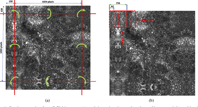
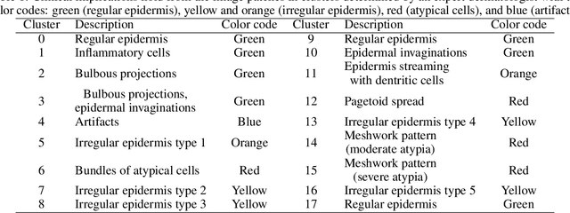
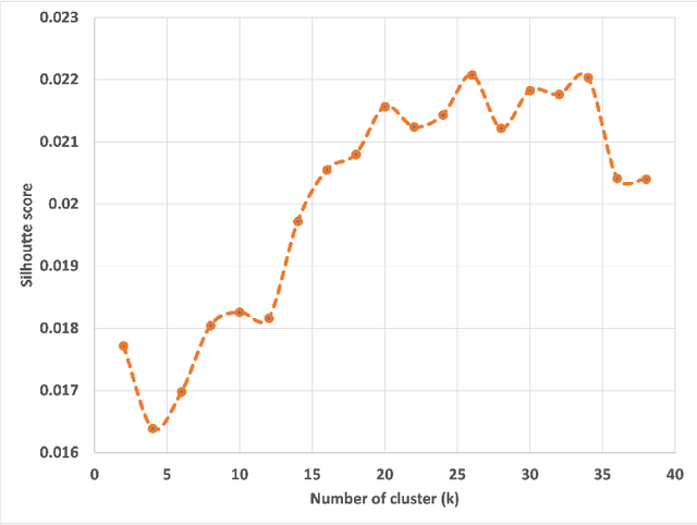
Abstract:Reflectance Confocal Microscopy (RCM) is a non-invasive imaging technique used in biomedical research and clinical dermatology. It provides virtual high-resolution images of the skin and superficial tissues, reducing the need for physical biopsies. RCM employs a laser light source to illuminate the tissue, capturing the reflected light to generate detailed images of microscopic structures at various depths. Recent studies explored AI and machine learning, particularly CNNs, for analyzing RCM images. Our study proposes a segmentation strategy based on textural features to identify clinically significant regions, empowering dermatologists in effective image interpretation and boosting diagnostic confidence. This approach promises to advance dermatological diagnosis and treatment.
 Add to Chrome
 Add to Firefox
 Add to Edge