Baljean Dhillon
A method for quantifying sectoral optic disc pallor in fundus photographs and its association with peripapillary RNFL thickness
Nov 13, 2023


Abstract:Purpose: To develop an automatic method of quantifying optic disc pallor in fundus photographs and determine associations with peripapillary retinal nerve fibre layer (pRNFL) thickness. Methods: We used deep learning to segment the optic disc, fovea, and vessels in fundus photographs, and measured pallor. We assessed the relationship between pallor and pRNFL thickness derived from optical coherence tomography scans in 118 participants. Separately, we used images diagnosed by clinical inspection as pale (N=45) and assessed how measurements compared to healthy controls (N=46). We also developed automatic rejection thresholds, and tested the software for robustness to camera type, image format, and resolution. Results: We developed software that automatically quantified disc pallor across several zones in fundus photographs. Pallor was associated with pRNFL thickness globally (\b{eta} = -9.81 (SE = 3.16), p < 0.05), in the temporal inferior zone (\b{eta} = -29.78 (SE = 8.32), p < 0.01), with the nasal/temporal ratio (\b{eta} = 0.88 (SE = 0.34), p < 0.05), and in the whole disc (\b{eta} = -8.22 (SE = 2.92), p < 0.05). Furthermore, pallor was significantly higher in the patient group. Lastly, we demonstrate the analysis to be robust to camera type, image format, and resolution. Conclusions: We developed software that automatically locates and quantifies disc pallor in fundus photographs and found associations between pallor measurements and pRNFL thickness. Translational relevance: We think our method will be useful for the identification, monitoring and progression of diseases characterized by disc pallor/optic atrophy, including glaucoma, compression, and potentially in neurodegenerative disorders.
Automated and Network Structure Preserving Segmentation of Optical Coherence Tomography Angiograms
Dec 20, 2019


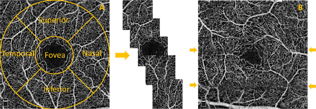
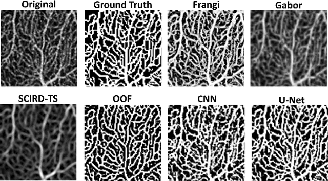
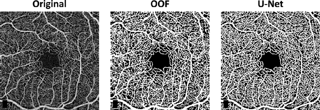
Abstract:Optical coherence tomography angiography (OCTA) is a novel non-invasive imaging modality for the visualisation of microvasculature in vivo. OCTA has encountered broad adoption in retinal research. OCTA potential in the assessment of pathological conditions and the reproducibility of studies relies on the quality of the image analysis. However, automated segmentation of parafoveal OCTA images is still an open problem in the field. In this study, we generate the first open dataset of retinal parafoveal OCTA images with associated ground truth manual segmentations. Furthermore, we establish a standard for OCTA image segmentation by surveying a broad range of state-of-the-art vessel enhancement and binarisation procedures. We provide the most comprehensive comparison of these methods under a unified framework to date. Our results show that, for the set of images considered, the U-Net machine learning (ML) architecture achieves the best performance with a Dice similarity coefficient of 0.89. For applications where manually segmented data is not available to retrain this ML approach, our findings suggest that optimal oriented flux is the best handcrafted filter enhancement method for OCTA images from those considered. Furthermore, we report on the importance of preserving network connectivity in the segmentation to enable vascular network phenotyping. We introduce a new metric for network connectivity evaluations in segmented angiograms and report an accuracy of up to 0.94 in preserving the morphological structure of the network in our segmentations. Finally, we release our data and source code to support standardisation efforts in OCTA image segmentation.
 Add to Chrome
 Add to Firefox
 Add to Edge