 Add to Chrome
 Add to Firefox
 Add to Edge
SLIM-Diff: Shared Latent Image-Mask Diffusion with Lp loss for Data-Scarce Epilepsy FLAIR MRI
Feb 03, 2026Focal cortical dysplasia (FCD) lesions in epilepsy FLAIR MRI are subtle and scarce, making joint image--mask generative modeling prone to instability and memorization. We propose SLIM-Diff, a compact joint diffusion model whose main contributions are (i) a single shared-bottleneck U-Net that enforces tight coupling between anatomy and lesion geometry from a 2-channel image+mask representation, and (ii) loss-geometry tuning via a tunable $L_p$ objective. As an internal baseline, we include the canonical DDPM-style objective ($ε$-prediction with $L_2$ loss) and isolate the effect of prediction parameterization and $L_p$ geometry under a matched setup. Experiments show that $x_0$-prediction is consistently the strongest choice for joint synthesis, and that fractional sub-quadratic penalties ($L_{1.5}$) improve image fidelity while $L_2$ better preserves lesion mask morphology. Our code and model weights are available in https://github.com/MarioPasc/slim-diff
Coronary Artery Disease Classification with Different Lesion Degree Ranges based on Deep Learning
Feb 01, 2024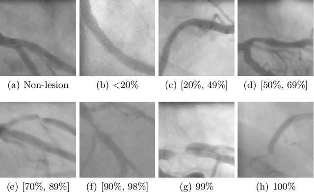
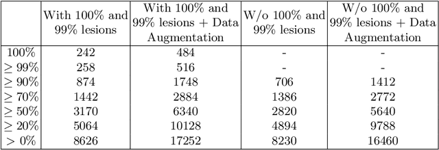
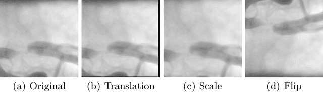
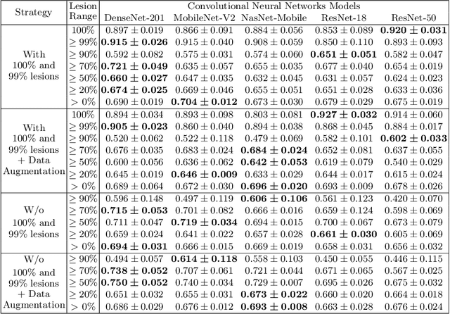
Invasive Coronary Angiography (ICA) images are considered the gold standard for assessing the state of the coronary arteries. Deep learning classification methods are widely used and well-developed in different areas where medical imaging evaluation has an essential impact due to the development of computer-aided diagnosis systems that can support physicians in their clinical procedures. In this paper, a new performance analysis of deep learning methods for binary ICA classification with different lesion degrees is reported. To reach this goal, an annotated dataset of ICA images that contains the ground truth, the location of lesions and seven possible severity degrees ranging between 0% and 100% was employed. The ICA images were divided into 'lesion' or 'non-lesion' patches. We aim to study how binary classification performance is affected by the different lesion degrees considered in the positive class. Therefore, five known convolutional neural network architectures were trained with different input images where different lesion degree ranges were gradually incorporated until considering the seven lesion degrees. Besides, four types of experiments with and without data augmentation were designed, whose F-measure and Area Under Curve (AUC) were computed. Reported results achieved an F-measure and AUC of 92.7% and 98.1%, respectively. However, lesion classification is highly affected by the degree of the lesion intended to classify, with 15% less accuracy when <99% lesion patches are present.
CADICA: a new dataset for coronary artery disease detection by using invasive coronary angiography
Feb 01, 2024Coronary artery disease (CAD) remains the leading cause of death globally and invasive coronary angiography (ICA) is considered the gold standard of anatomical imaging evaluation when CAD is suspected. However, risk evaluation based on ICA has several limitations, such as visual assessment of stenosis severity, which has significant interobserver variability. This motivates to development of a lesion classification system that can support specialists in their clinical procedures. Although deep learning classification methods are well-developed in other areas of medical imaging, ICA image classification is still at an early stage. One of the most important reasons is the lack of available and high-quality open-access datasets. In this paper, we reported a new annotated ICA images dataset, CADICA, to provide the research community with a comprehensive and rigorous dataset of coronary angiography consisting of a set of acquired patient videos and associated disease-related metadata. This dataset can be used by clinicians to train their skills in angiographic assessment of CAD severity and by computer scientists to create computer-aided diagnostic systems to help in such assessment. In addition, baseline classification methods are proposed and analyzed, validating the functionality of CADICA and giving the scientific community a starting point to improve CAD detection.