Anja Zenz
Self-Supervised Learning from Unlabeled Fundus Photographs Improves Segmentation of the Retina
Aug 05, 2021


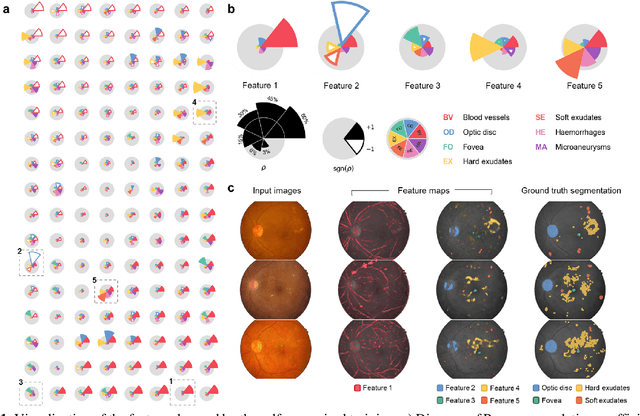
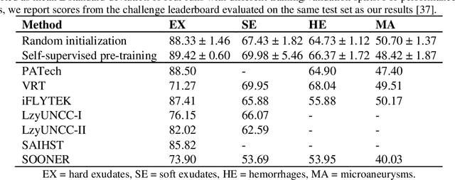
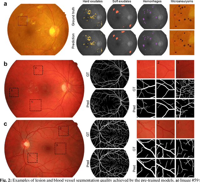
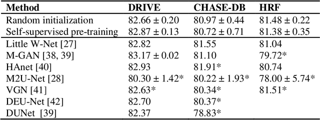
Abstract:Fundus photography is the primary method for retinal imaging and essential for diabetic retinopathy prevention. Automated segmentation of fundus photographs would improve the quality, capacity, and cost-effectiveness of eye care screening programs. However, current segmentation methods are not robust towards the diversity in imaging conditions and pathologies typical for real-world clinical applications. To overcome these limitations, we utilized contrastive self-supervised learning to exploit the large variety of unlabeled fundus images in the publicly available EyePACS dataset. We pre-trained an encoder of a U-Net, which we later fine-tuned on several retinal vessel and lesion segmentation datasets. We demonstrate for the first time that by using contrastive self-supervised learning, the pre-trained network can recognize blood vessels, optic disc, fovea, and various lesions without being provided any labels. Furthermore, when fine-tuned on a downstream blood vessel segmentation task, such pre-trained networks achieve state-of-the-art performance on images from different datasets. Additionally, the pre-training also leads to shorter training times and an improved few-shot performance on both blood vessel and lesion segmentation tasks. Altogether, our results showcase the benefits of contrastive self-supervised pre-training which can play a crucial role in real-world clinical applications requiring robust models able to adapt to new devices with only a few annotated samples.
 Add to Chrome
 Add to Firefox
 Add to Edge