Amin Komeili
Swin UNETR segmentation with automated geometry filtering for biomechanical modeling of knee joint cartilage
Jul 08, 2024


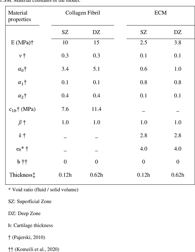
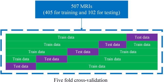
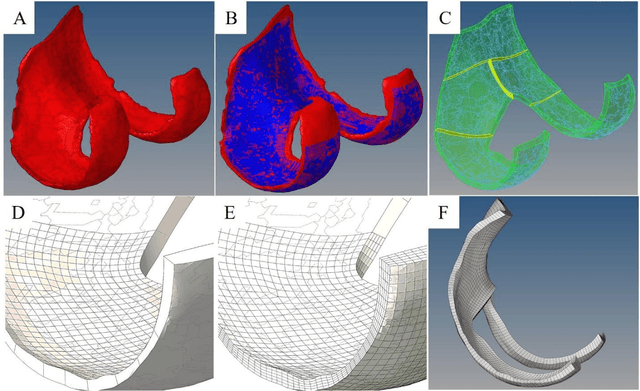
Abstract:Simulation studies, such as finite element (FE) modeling, offer insights into knee joint biomechanics, which may not be achieved through experimental methods without direct involvement of patients. While generic FE models have been used to predict tissue biomechanics, they overlook variations in population-specific geometry, loading, and material properties. In contrast, subject-specific models account for these factors, delivering enhanced predictive precision but requiring significant effort and time for development. This study aimed to facilitate subject-specific knee joint FE modeling by integrating an automated cartilage segmentation algorithm using a 3D Swin UNETR. This algorithm provided initial segmentation of knee cartilage, followed by automated geometry filtering to refine surface roughness and continuity. In addition to the standard metrics of image segmentation performance, such as Dice similarity coefficient (DSC) and Hausdorff distance, the method's effectiveness was also assessed in FE simulation. Nine pairs of knee cartilage FE models, using manual and automated segmentation methods, were developed to compare the predicted stress and strain responses during gait. The automated segmentation achieved high Dice similarity coefficients of 89.4% for femoral and 85.1% for tibial cartilage, with a Hausdorff distance of 2.3 mm between the automated and manual segmentation. Mechanical results including maximum principal stress and strain, fluid pressure, fibril strain, and contact area showed no significant differences between the manual and automated FE models. These findings demonstrate the effectiveness of the proposed automated segmentation method in creating accurate knee joint FE models.
Automatic Classification of Symmetry of Hemithoraces in Canine and Feline Radiographs
Feb 24, 2023Abstract:Purpose: Thoracic radiographs are commonly used to evaluate patients with confirmed or suspected thoracic pathology. Proper patient positioning is more challenging in canine and feline radiography than in humans due to less patient cooperation and body shape variation. Improper patient positioning during radiograph acquisition has the potential to lead to a misdiagnosis. Asymmetrical hemithoraces are one of the indications of obliquity for which we propose an automatic classification method. Approach: We propose a hemithoraces segmentation method based on Convolutional Neural Networks (CNNs) and active contours. We utilized the U-Net model to segment the ribs and spine and then utilized active contours to find left and right hemithoraces. We then extracted features from the left and right hemithoraces to train an ensemble classifier which includes Support Vector Machine, Gradient Boosting and Multi-Layer Perceptron. Five-fold cross-validation was used, thorax segmentation was evaluated by Intersection over Union (IoU), and symmetry classification was evaluated using Precision, Recall, Area under Curve and F1 score. Results: Classification of symmetry for 900 radiographs reported an F1 score of 82.8% . To test the robustness of the proposed thorax segmentation method to underexposure and overexposure, we synthetically corrupted properly exposed radiographs and evaluated results using IoU. The results showed that the models IoU for underexposure and overexposure dropped by 2.1% and 1.2%, respectively. Conclusions: Our results indicate that the proposed thorax segmentation method is robust to poor exposure radiographs. The proposed thorax segmentation method can be applied to human radiography with minimal changes.
 Add to Chrome
 Add to Firefox
 Add to Edge