Aman Swaraj
Classification of COVID-19 Patients with their Severity Level from Chest CT Scans using Transfer Learning
May 27, 2022
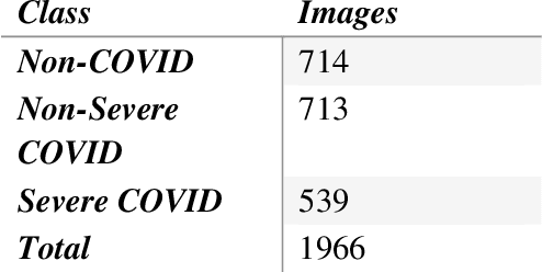
Abstract:Background and Objective: During pandemics, the use of artificial intelligence (AI) approaches combined with biomedical science play a significant role in reducing the burden on the healthcare systems and physicians. The rapid increment in cases of COVID-19 has led to an increase in demand for hospital beds and other medical equipment. However, since medical facilities are limited, it is recommended to diagnose patients as per the severity of the infection. Keeping this in mind, we share our research in detecting COVID-19 as well as assessing its severity using chest-CT scans and Deep Learning pre-trained models. Dataset: We have collected a total of 1966 CT Scan images for three different class labels, namely, Non-COVID, Severe COVID, and Non-Severe COVID, out of which 714 CT images belong to the Non-COVID category, 713 CT images are for Non-Severe COVID category and 539 CT images are of Severe COVID category. Methods: All of the images are initially pre-processed using the Contrast Limited Histogram Equalization (CLAHE) approach. The pre-processed images are then fed into the VGG-16 network for extracting features. Finally, the retrieved characteristics are categorized and the accuracy is evaluated using a support vector machine (SVM) with 10-fold cross-validation (CV). Result and Conclusion: In our study, we have combined well-known strategies for pre-processing, feature extraction, and classification which brings us to a remarkable success rate of disease and its severity recognition with an accuracy of 96.05% (97.7% for Non-Severe COVID-19 images and 93% for Severe COVID-19 images). Our model can therefore help radiologists detect COVID-19 and the extent of its severity.
COVID-19 Severity Classification on Chest X-ray Images
May 25, 2022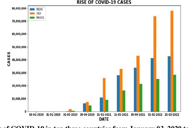

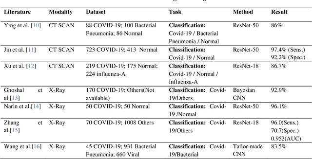
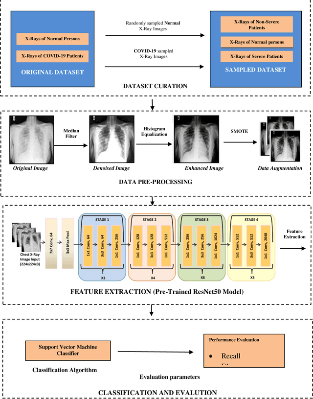
Abstract:Biomedical imaging analysis combined with artificial intelligence (AI) methods has proven to be quite valuable in order to diagnose COVID-19. So far, various classification models have been used for diagnosing COVID-19. However, classification of patients based on their severity level is not yet analyzed. In this work, we classify covid images based on the severity of the infection. First, we pre-process the X-ray images using a median filter and histogram equalization. Enhanced X-ray images are then augmented using SMOTE technique for achieving a balanced dataset. Pre-trained Resnet50, VGG16 model and SVM classifier are then used for feature extraction and classification. The result of the classification model confirms that compared with the alternatives, with chest X-Ray images, the ResNet-50 model produced remarkable classification results in terms of accuracy (95%), recall (0.94), and F1-Score (0.92), and precision (0.91).
Classification of COVID-19 on chest X-Ray images using Deep Learning model with Histogram Equalization and Lungs Segmentation
Dec 05, 2021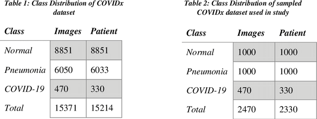
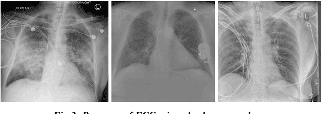
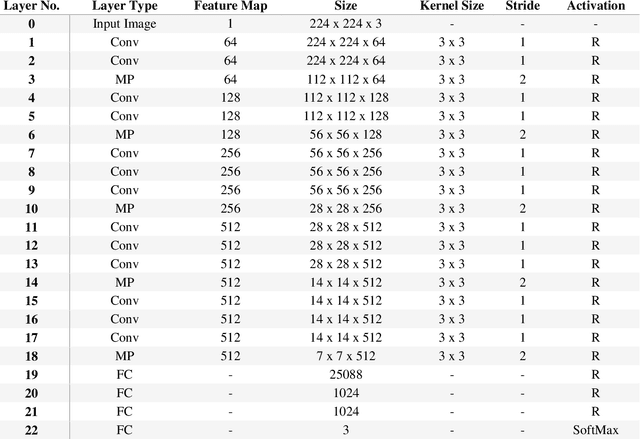
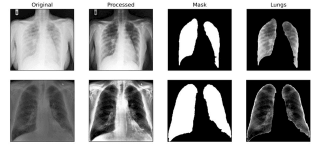
Abstract:Background and Objective: Artificial intelligence (AI) methods coupled with biomedical analysis has a critical role during pandemics as it helps to release the overwhelming pressure from healthcare systems and physicians. As the ongoing COVID-19 crisis worsens in countries having dense populations and inadequate testing kits like Brazil and India, radiological imaging can act as an important diagnostic tool to accurately classify covid-19 patients and prescribe the necessary treatment in due time. With this motivation, we present our study based on deep learning architecture for detecting covid-19 infected lungs using chest X-rays. Dataset: We collected a total of 2470 images for three different class labels, namely, healthy lungs, ordinary pneumonia, and covid-19 infected pneumonia, out of which 470 X-ray images belong to the covid-19 category. Methods: We first pre-process all the images using histogram equalization techniques and segment them using U-net architecture. VGG-16 network is then used for feature extraction from the pre-processed images which is further sampled by SMOTE oversampling technique to achieve a balanced dataset. Finally, the class-balanced features are classified using a support vector machine (SVM) classifier with 10-fold cross-validation and the accuracy is evaluated. Result and Conclusion: Our novel approach combining well-known pre-processing techniques, feature extraction methods, and dataset balancing method, lead us to an outstanding rate of recognition of 98% for COVID-19 images over a dataset of 2470 X-ray images. Our model is therefore fit to be utilized in healthcare facilities for screening purposes.
Comparison of Traditional and Hybrid Time Series Models for Forecasting COVID-19 Cases
May 05, 2021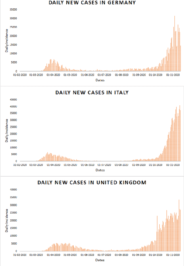
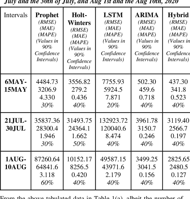
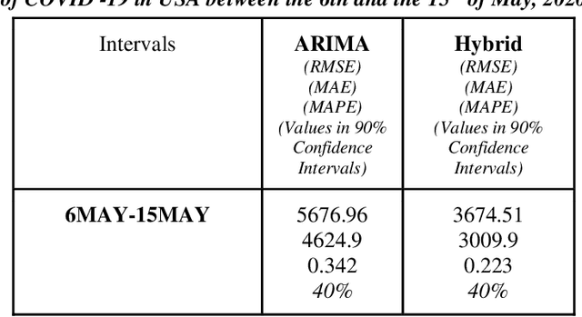

Abstract:Time series forecasting methods play critical role in estimating the spread of an epidemic. The coronavirus outbreak of December 2019 has already infected millions all over the world and continues to spread on. Just when the curve of the outbreak had started to flatten, many countries have again started to witness a rise in cases which is now being referred as the 2nd wave of the pandemic. A thorough analysis of time-series forecasting models is therefore required to equip state authorities and health officials with immediate strategies for future times. This aims of the study are three-fold: (a) To model the overall trend of the spread; (b) To generate a short-term forecast of 10 days in countries with the highest incidence of confirmed cases (USA, India and Brazil); (c) To quantitatively determine the algorithm that is best suited for precise modelling of the linear and non-linear features of the time series. The comparison of forecasting models for the total cumulative cases of each country is carried out by comparing the reported data and the predicted value, and then ranking the algorithms (Prophet, Holt-Winters, LSTM, ARIMA, and ARIMA-NARNN) based on their RMSE, MAE and MAPE values. The hybrid combination of ARIMA and NARNN (Nonlinear Auto-Regression Neural Network) gave the best result among the selected models with a reduced RMSE, which proved to be almost 35.3% better than one of the most prevalent method of time-series prediction (ARIMA). The results demonstrated the efficacy of the hybrid implementation of the ARIMA-NARNN model over other forecasting methods such as Prophet, Holt Winters, LSTM, and the ARIMA model in encapsulating the linear as well as non-linear patterns of the epidemical datasets.
 Add to Chrome
 Add to Firefox
 Add to Edge