Alexandre Fioravante de Siqueira
A reusable pipeline for large-scale fiber segmentation on unidirectional fiber beds using fully convolutional neural networks
Jan 15, 2021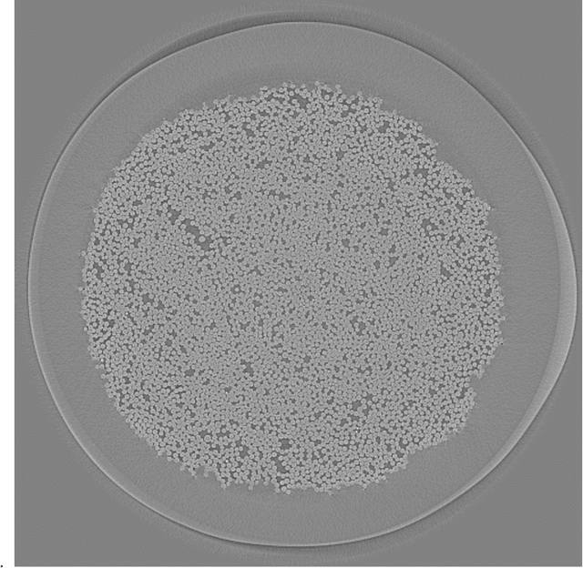

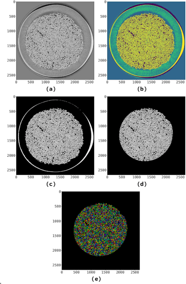
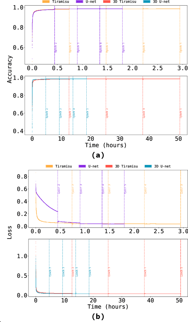
Abstract:Fiber-reinforced ceramic-matrix composites are advanced materials resistant to high temperatures, with application to aerospace engineering. Their analysis depends on the detection of embedded fibers, with semi-supervised techniques usually employed to separate fibers within the fiber beds. Here we present an open computational pipeline to detect fibers in ex-situ X-ray computed tomography fiber beds. To separate the fibers in these samples, we tested four different architectures of fully convolutional neural networks. When comparing our neural network approach to a semi-supervised one, we obtained Dice and Matthews coefficients greater than $92.28 \pm 9.65\%$, reaching up to $98.42 \pm 0.03 \%$, showing that the network results are close to the human-supervised ones in these fiber beds, in some cases separating fibers that human-curated algorithms could not find. The software we generated in this project is open source, released under a permissive license, and can be freely adapted and re-used in other domains. All data and instructions on how to download and use it are also available.
Automatic counting of fission tracks in apatite and muscovite using image processing
Jun 13, 2018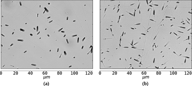

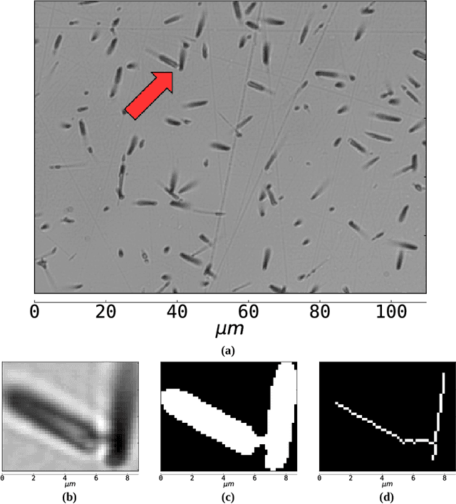
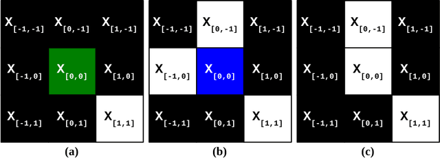
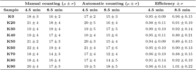
Abstract:One of the major difficulties of automatic track counting using photomicrographs is separating overlapped tracks. We address this issue combining image processing algorithms such as skeletonization, and we test our algorithm with several binarization techniques. The counting algorithm was successfully applied to determine the efficiency factor GQR, necessary for standardless fission-track dating, involving counting induced tracks in apatite and muscovite with superficial densities of about $6 \times 10^5$ tracks/$cm^2$.
Segmentation of nearly isotropic overlapped tracks in photomicrographs using successive erosions as watershed markers
Jan 18, 2018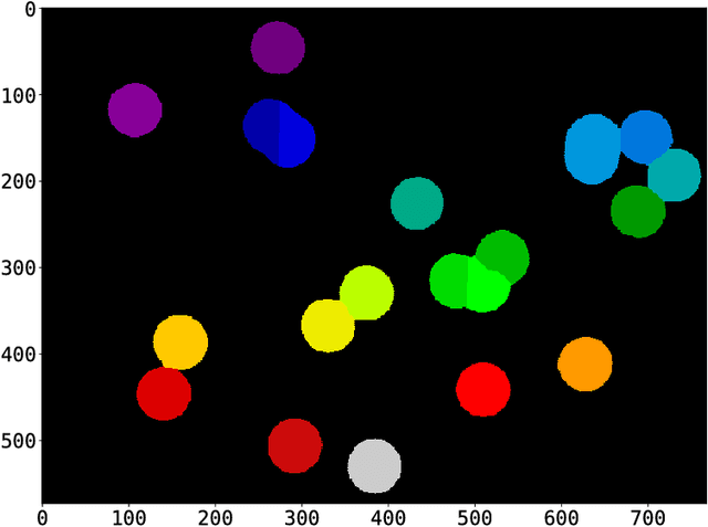
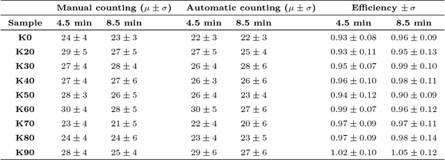
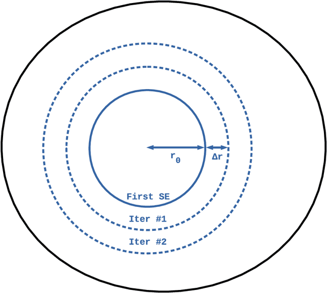
Abstract:The major challenges of automatic track counting are distinguishing tracks and material defects, identifying small tracks and defects of similar size, and detecting overlapping tracks. Here we address the latter issue using WUSEM, an algorithm which combines the watershed transform, morphological erosions and labeling to separate regions in photomicrographs. WUSEM shows reliable results when used in photomicrographs presenting almost isotropic objects. We tested this method in two datasets of diallyl phthalate (DAP) photomicrographs and compared the results when counting manually and using the classic watershed. The mean automatic/manual efficiency ratio when using WUSEM in the test datasets is 0.97 +/- 0.11.
Estimating the concentration of gold nanoparticles incorporated on Natural Rubber membranes using Multi-Level Starlet Optimal Segmentation
Oct 26, 2016


Abstract:This study consolidates Multi-Level Starlet Segmentation (MLSS) and Multi-Level Starlet Optimal Segmentation (MLSOS), techniques for photomicrograph segmentation that use starlet wavelet detail levels to separate areas of interest in an input image. Several segmentation levels can be obtained using Multi-Level Starlet Segmentation; after that, Matthews correlation coefficient (MCC) is used to choose an optimal segmentation level, giving rise to Multi-Level Starlet Optimal Segmentation. In this paper, MLSOS is employed to estimate the concentration of gold nanoparticles with diameter around 47 nm, reducted on natural rubber membranes. These samples were used on the construction of SERS/SERRS substrates and in the study of natural rubber membranes with incorporated gold nanoparticles influence on Leishmania braziliensis physiology. Precision, recall and accuracy are used to evaluate the segmentation performance, and MLSOS presents accuracy greater than 88% for this application.
* 22 pages, 8 figures
Segmentation of scanning electron microscopy images from natural rubber samples with gold nanoparticles using starlet wavelets
Jun 12, 2016


Abstract:Electronic microscopy has been used for morphology evaluation of different materials structures. However, microscopy results may be affected by several factors. Image processing methods can be used to correct and improve the quality of these results. In this paper we propose an algorithm based on starlets to perform the segmentation of scanning electron microscopy images. An application is presented in order to locate gold nanoparticles in natural rubber membranes. In this application, our method showed accuracy greater than 85% for all test images. Results given by this method will be used in future studies, to computationally estimate the density distribution of gold nanoparticles in natural rubber samples and to predict reduction kinetics of gold nanoparticles at different time periods.
Jansen-MIDAS: a multi-level photomicrograph segmentation software based on isotropic undecimated wavelets
May 25, 2016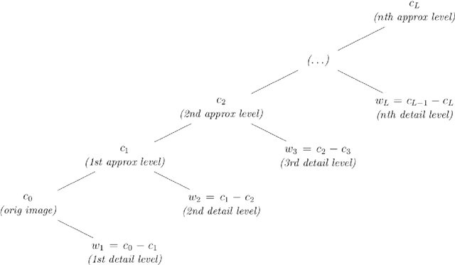
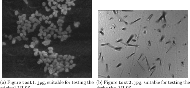
Abstract:Image segmentation, the process of separating the elements within an image, is frequently used for obtaining information from photomicrographs. However, segmentation methods should be used with reservations: incorrect segmentation can mislead when interpreting regions of interest (ROI), thus decreasing the success rate of additional procedures. Multi-Level Starlet Segmentation (MLSS) and Multi-Level Starlet Optimal Segmentation (MLSOS) were developed to address the photomicrograph segmentation deficiency on general tools. These methods gave rise to Jansen-MIDAS, an open-source software which a scientist can use to obtain a multi-level threshold segmentation of his/hers photomicrographs. This software is presented in two versions: a text-based version, for GNU Octave, and a graphical user interface (GUI) version, for MathWorks MATLAB. It can be used to process several types of images, becoming a reliable alternative to the scientist.
An automatic method for segmentation of fission tracks in epidote crystal photomicrographs
Feb 12, 2016


Abstract:Manual identification of fission tracks has practical problems, such as variation due to observer-observation efficiency. An automatic processing method that could identify fission tracks in a photomicrograph could solve this problem and improve the speed of track counting. However, separation of non-trivial images is one of the most difficult tasks in image processing. Several commercial and free softwares are available, but these softwares are meant to be used in specific images. In this paper, an automatic method based on starlet wavelets is presented in order to separate fission tracks in mineral photomicrographs. Automatization is obtained by Matthews correlation coefficient, and results are evaluated by precision, recall and accuracy. This technique is an improvement of a method aimed at segmentation of scanning electron microscopy images. This method is applied in photomicrographs of epidote phenocrystals, in which accuracy higher than 89% was obtained in fission track segmentation, even for difficult images. Algorithms corresponding to the proposed method are available for download. Using the method presented here, an user could easily determine fission tracks in photomicrographs of mineral samples.
* 16 pages, 5 figures
 Add to Chrome
 Add to Firefox
 Add to Edge