Tissue segmentation with deep 3D networks and spatial priors
Paper and Code
May 24, 2019


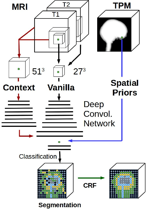
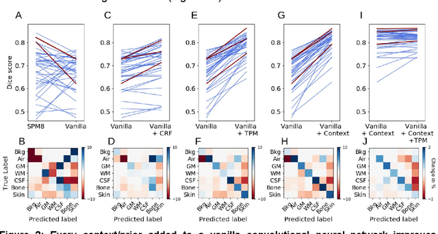
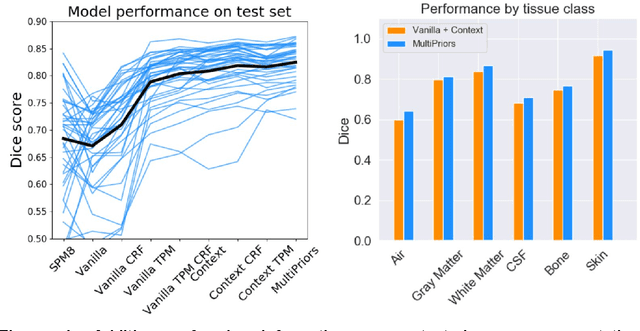
Conventional automated segmentation of the human head distinguishes different tissues based on image intensities in an MRI volume and prior tissue probability maps (TPM). This works well for normal head anatomies, but fails in the presence of unexpected lesions. Deep convolutional neural networks leverage instead volumetric spatial patterns and can be trained to segment lesions, but have thus far not integrated prior probabilities. Here we add to a three-dimensional convolutional network spatial priors with a TPM, morphological priors with conditional random fields, and context with a wider field-of-view at lower resolution. The new architecture, which we call MultiPrior, was designed to be a fully-trainable, three-dimensional convolutional network. Thus, the resulting architecture represents a neural network with learnable spatial memories. When trained on a set of stroke patients and healthy subjects, MultiPrior outperforms the state-of-the-art segmentation tools such as DeepMedic and SPM segmentation. The approach is further demonstrated on patients with disorders of consciousness, where we find that cognitive state correlates positively with gray-matter volumes and negatively with the extent of ventricles. We make the code and trained networks freely available to support future clinical research projects.
 Add to Chrome
 Add to Firefox
 Add to Edge