Spatial response identification enables robust experimental ultrasound computed tomography
Paper and Code
Apr 05, 2021


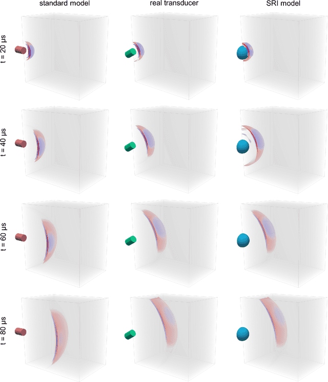
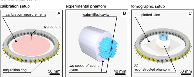
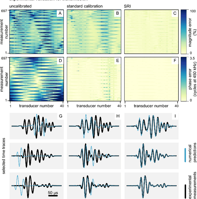
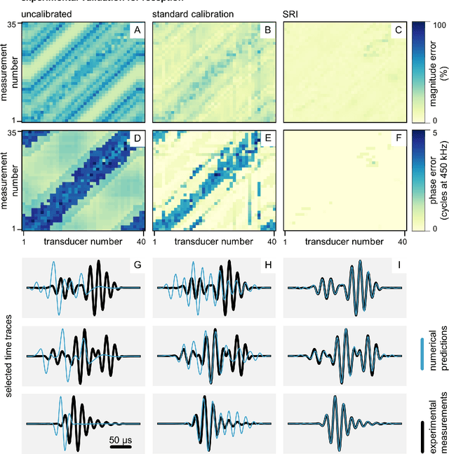
Ultrasound computed tomography techniques have the potential to provide clinicians with 3D, quantitative and high-resolution information of both soft and hard tissues such as the breast or the adult human brain. Their practical application requires accurate modelling of the acquisition setup: the spatial location, orientation, and impulse response of each ultrasound transducer. However, existing calibration methods fail to accurately characterise these transducers unless their size can be considered negligible when compared to the dominant wavelength, which reduces signal-to-noise ratios below usable levels in the presence of high-contrast tissues such as the skull. In this paper, we introduce a methodology that can simultaneously estimate the location, orientation, and impulse response of the ultrasound transducers in a single calibration. We do this by extending spatial response identification, an algorithm that we have recently proposed to estimate transducer impulse responses. Our proposed methodology replaces the transducers in the acquisition device with a surrogate model whose effective response matches the experimental data by fitting a numerical model of wave propagation. This results in a flexible and robust calibration procedure that can accurately predict the behaviour of the ultrasound acquisition device without ever having to know where the real transducers are or their individual impulse response. Experimental results using a ring acquisition system show that spatial response identification produces calibrations of significantly higher quality than standard methodologies across all transducers, both in transmission and in reception. Experimental full-waveform inversion reconstructions of a tissue-mimicking phantom demonstrate that spatial response identification generates more accurate reconstructions than those produced with standard calibration techniques.
 Add to Chrome
 Add to Firefox
 Add to Edge