Self-Training Based Unsupervised Cross-Modality Domain Adaptation for Vestibular Schwannoma and Cochlea Segmentation
Paper and Code
Sep 22, 2021


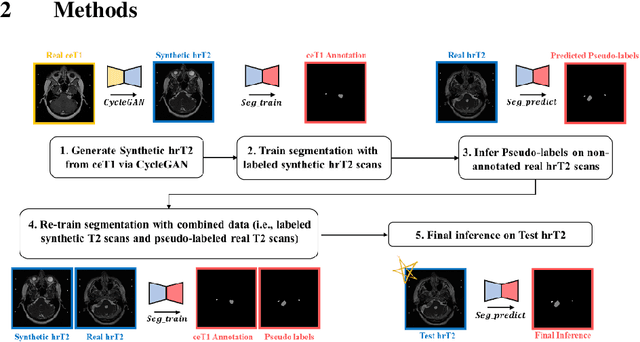
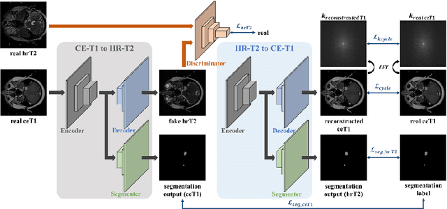
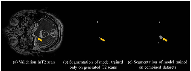
With the advances of deep learning, many medical image segmentation studies achieve human-level performance when in fully supervised condition. However, it is extremely expensive to acquire annotation on every data in medical fields, especially on magnetic resonance images (MRI) that comprise many different contrasts. Unsupervised methods can alleviate this problem; however, the performance drop is inevitable compared to fully supervised methods. In this work, we propose a self-training based unsupervised-learning framework that performs automatic segmentation of Vestibular Schwannoma (VS) and cochlea on high-resolution T2 scans. Our method consists of 4 main stages: 1) VS-preserving contrast conversion from contrast-enhanced T1 scan to high-resolution T2 scan, 2) training segmentation on generated T2 scans with annotations on T1 scans, and 3) Inferring pseudo-labels on non-annotated real T2 scans, and 4) boosting the generalizability of VS and cochlea segmentation by training with combined data (i.e., real T2 scans with pseudo-labels and generated T2 scans with true annotations). Our method showed mean Dice score and Average Symmetric Surface Distance (ASSD) of 0.8570 (0.0705) and 0.4970 (0.3391) for VS, 0.8446 (0.0211) and 0.1513 (0.0314) for Cochlea on CrossMoDA2021 challenge validation phase leaderboard, outperforming most other approaches.
 Add to Chrome
 Add to Firefox
 Add to Edge