Dynamic optical contrast imaging for real-time delineation of tumor resection margins using head and neck cancer as a model
Paper and Code
Mar 10, 2022


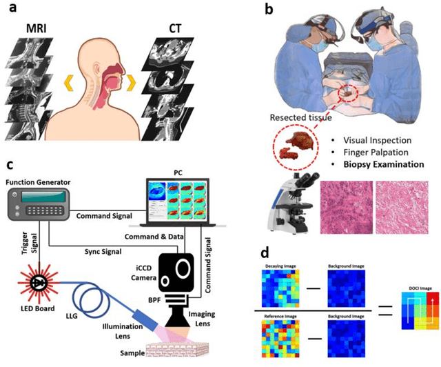
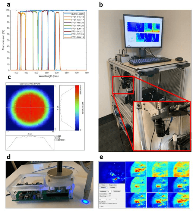
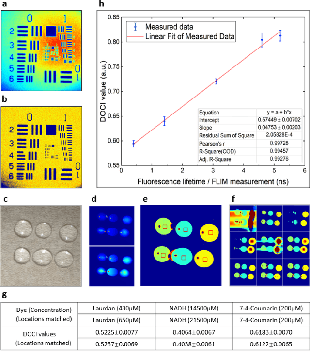
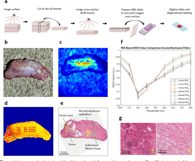
Complete surgical resection of the tumor for Head and neck squamous cell carcinoma (HNSCC) remains challenging, given the devastating side effects of aggressive surgery and the anatomic proximity to vital structures. To address the clinical challenges, we introduce a wide-field, label-free imaging tool that can assist surgeons delineate tumor margins real-time. We assume that autofluorescence lifetime is a natural indicator of the health level of tissues, and ratio-metric measurement of the emission-decay state to the emission-peak state of excited fluorophores will enable rapid lifetime mapping of tissues. Here, we describe the principle, instrumentation, characterization of the imager and the intraoperative imaging of resected tissues from 13 patients undergoing head and neck cancer resection. 20 x 20 mm2 imaging takes 2 second/frame with a working distance of 50 mm, and characterization shows that the spatial resolution reached 70 {\mu}m and the least distinguishable fluorescence lifetime difference is 0.14 ns. Tissue imaging and Hematoxylin-Eosin stain slides comparison reveals its capability of delineating cancerous boundaries with submillimeter accuracy and a sensitivity of 91.86% and specificity of 84.38%.
 Add to Chrome
 Add to Firefox
 Add to Edge