Deep Neural Networks for Anatomical Brain Segmentation
Paper and Code
Jun 25, 2015


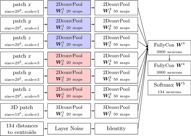
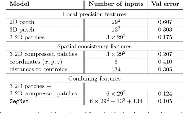
We present a novel approach to automatically segment magnetic resonance (MR) images of the human brain into anatomical regions. Our methodology is based on a deep artificial neural network that assigns each voxel in an MR image of the brain to its corresponding anatomical region. The inputs of the network capture information at different scales around the voxel of interest: 3D and orthogonal 2D intensity patches capture the local spatial context while large, compressed 2D orthogonal patches and distances to the regional centroids enforce global spatial consistency. Contrary to commonly used segmentation methods, our technique does not require any non-linear registration of the MR images. To benchmark our model, we used the dataset provided for the MICCAI 2012 challenge on multi-atlas labelling, which consists of 35 manually segmented MR images of the brain. We obtained competitive results (mean dice coefficient 0.725, error rate 0.163) showing the potential of our approach. To our knowledge, our technique is the first to tackle the anatomical segmentation of the whole brain using deep neural networks.
 Add to Chrome
 Add to Firefox
 Add to Edge