A System for 3D Reconstruction Of Comminuted Tibial Plafond Bone Fractures
Paper and Code
Feb 23, 2021


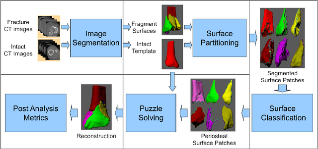
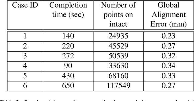
High energy impacts at joint locations often generate highly fragmented, or comminuted, bone fractures. Current approaches for treatment require physicians to decide how to classify the fracture within a hierarchy fracture severity categories. Each category then provides a best-practice treatment scenario to obtain the best possible prognosis for the patient. This article identifies shortcomings associated with qualitative-only evaluation of fracture severity and provides new quantitative metrics that serve to address these shortcomings. We propose a system to semi-automatically extract quantitative metrics that are major indicators of fracture severity. These include: (i) fracture surface area, i.e., how much surface area was generated when the bone broke apart, and (ii) dispersion, i.e., how far the fragments have rotated and translated from their original anatomic positions. This article describes new computational tools to extract these metrics by computationally reconstructing 3D bone anatomy from CT images with a focus on tibial plafond fracture cases where difficult qualitative fracture severity cases are more prevalent. Reconstruction is accomplished within a single system that integrates several novel algorithms that identify, extract and piece-together fractured fragments in a virtual environment. Doing so provides objective quantitative measures for these fracture severity indicators. The availability of such measures provides new tools for fracture severity assessment which may lead to improved fracture treatment. This paper describes the system, the underlying algorithms and the metrics of the reconstruction results by quantitatively analyzing six clinical tibial plafond fracture cases.
 Add to Chrome
 Add to Firefox
 Add to Edge