Upender Kalwa
An automated, high-resolution phenotypic assay for adult Brugia malayi and microfilaria
Sep 05, 2023Abstract:Brugia malayi are thread-like parasitic worms and one of the etiological agents of Lymphatic filariasis (LF). Existing anthelmintic drugs to treat LF are effective in reducing the larval microfilaria (mf) counts in human bloodstream but are less effective on adult parasites. To test potential drug candidates, we report a multi-parameter phenotypic assay based on tracking the motility of adult B. malayi and mf in vitro. For adult B. malayi, motility is characterized by the centroid velocity, path curvature, angular velocity, eccentricity, extent, and Euler Number. These parameters are evaluated in experiments with three anthelmintic drugs. For B. malayi mf, motility is extracted from the evolving body skeleton to yield positional data and bending angles at 74 key point. We achieved high-fidelity tracking of complex worm postures (self-occlusions, omega turns, body bending, and reversals) while providing a visual representation of pose estimates and behavioral attributes in both space and time scales.
Flexible and disposable paper- and plastic-based gel micropads for nematode handling, imaging, and chemical testing
Jun 23, 2022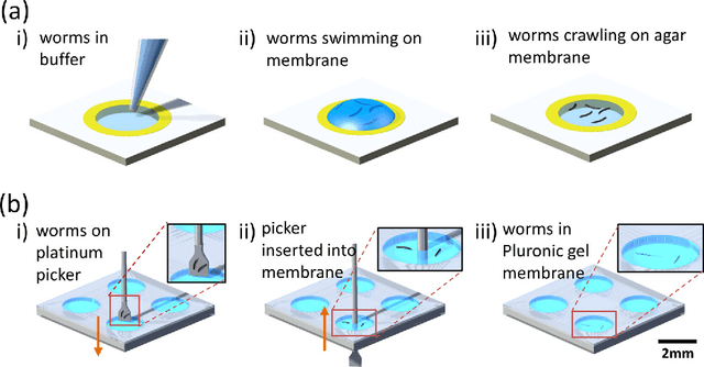
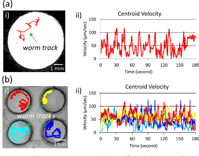

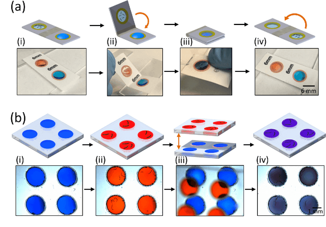
Abstract:Today, the area of point-of-care diagnostics is synonymous with paper microfluidics where cheap, disposable, and on-the-spot detection toolkits are being developed for a variety of chemical tests. In this work, we present a novel application of microfluidic paper-based analytical devices (microPADs) to study the behavior of a small model nematode, Caenorhabditis elegans. We describe schemes of microPAD fabrication on paper and plastic substrates where membranes are created in agarose and Pluronic gel. Methods are demonstrated for loading, visualizing, and transferring single and multiple nematodes. Using an anthelmintic drug, levamisole, we show that chemical testing on C. elegans is easily performed because of the open device structure. A custom program is written to automatically recognize individual worms on the microPADs and extract locomotion parameters in real-time. The combination of microPADs and the nematode tracking program provides a relatively low-cost, simple-to-fabricate imaging and screening assay (compared to standard agarose plates or polymeric microfluidic devices) for non-microfluidic, nematode laboratories.
Skin Cancer Diagnostics with an All-Inclusive Smartphone Application
May 25, 2022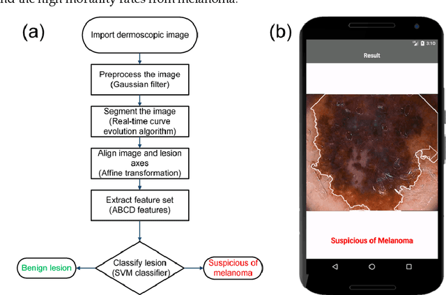
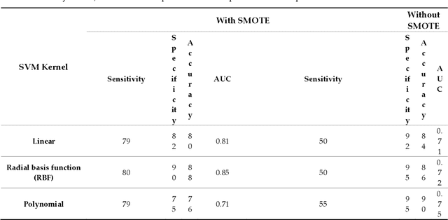
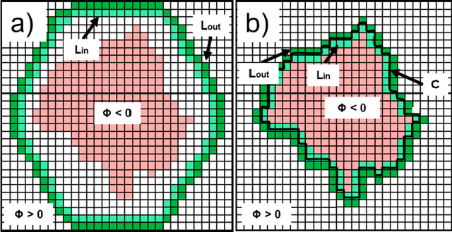

Abstract:Among the different types of skin cancer, melanoma is considered to be the deadliest and is difficult to treat at advanced stages. Detection of melanoma at earlier stages can lead to reduced mortality rates. Desktop-based computer-aided systems have been developed to assist dermatologists with early diagnosis. However, there is significant interest in developing portable, at-home melanoma diagnostic systems which can assess the risk of cancerous skin lesions. Here, we present a smartphone application that combines image capture capabilities with preprocessing and segmentation to extract the Asymmetry, Border irregularity, Color variegation, and Diameter (ABCD) features of a skin lesion. Using the feature sets, classification of malignancy is achieved through support vector machine classifiers. By using adaptive algorithms in the individual data-processing stages, our approach is made computationally light, user friendly, and reliable in discriminating melanoma cases from benign ones. Images of skin lesions are either captured with the smartphone camera or imported from public datasets. The entire process from image capture to classification runs on an Android smartphone equipped with a detachable 10x lens, and processes an image in less than a second. The overall performance metrics are evaluated on a public database of 200 images with Synthetic Minority Over-sampling Technique (SMOTE) (80% sensitivity, 90% specificity, 88% accuracy, and 0.85 area under curve (AUC)) and without SMOTE (55% sensitivity, 95% specificity, 90% accuracy, and 0.75 AUC). The evaluated performance metrics and computation times are comparable or better than previous methods. This all-inclusive smartphone application is designed to be easy-to-download and easy-to-navigate for the end user, which is imperative for the eventual democratization of such medical diagnostic systems.
New methods of removing debris and high-throughput counting of cyst nematode eggs extracted from field soil
May 25, 2022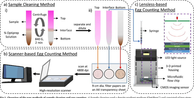
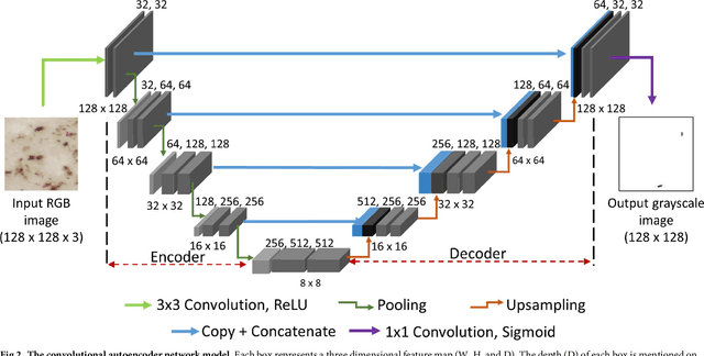
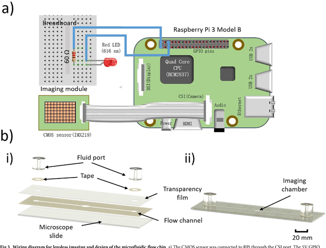
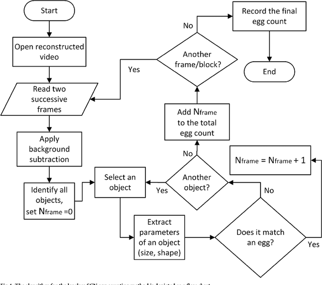
Abstract:The soybean cyst nematode (SCN), Heterodera glycines, is the most damaging pathogen of soybeans in the United States. To assess the severity of nematode infestations in the field, SCN egg population densities are determined. Cysts (dead females) of the nematode must be extracted from soil samples and then ground to extract the eggs within. Sucrose centrifugation commonly is used to separate debris from suspensions of extracted nematode eggs. We present a method using OptiPrep as a density gradient medium with improved separation and recovery of extracted eggs compared to the sucrose centrifugation technique. Also, computerized methods were developed to automate the identification and counting of nematode eggs from the processed samples. In one approach, a high-resolution scanner was used to take static images of extracted eggs and debris on filter papers, and a deep learning network was trained to identify and count the eggs among the debris. In the second approach, a lensless imaging setup was developed using off-the-shelf components, and the processed egg samples were passed through a microfluidic flow chip made from double-sided adhesive tape. Holographic videos were recorded of the passing eggs and debris, and the videos were reconstructed and processed by custom software program to obtain egg counts. The performance of the software programs for egg counting was characterized with SCN-infested soil collected from two farms, and the results using these methods were compared with those obtained through manual counting.
 Add to Chrome
 Add to Firefox
 Add to Edge