 Add to Chrome
 Add to Firefox
 Add to Edge
3D Foundation AI Model for Generalizable Disease Detection in Head Computed Tomography
Feb 04, 2025


Head computed tomography (CT) imaging is a widely-used imaging modality with multitudes of medical indications, particularly in assessing pathology of the brain, skull, and cerebrovascular system. It is commonly the first-line imaging in neurologic emergencies given its rapidity of image acquisition, safety, cost, and ubiquity. Deep learning models may facilitate detection of a wide range of diseases. However, the scarcity of high-quality labels and annotations, particularly among less common conditions, significantly hinders the development of powerful models. To address this challenge, we introduce FM-CT: a Foundation Model for Head CT for generalizable disease detection, trained using self-supervised learning. Our approach pre-trains a deep learning model on a large, diverse dataset of 361,663 non-contrast 3D head CT scans without the need for manual annotations, enabling the model to learn robust, generalizable features. To investigate the potential of self-supervised learning in head CT, we employed both discrimination with self-distillation and masked image modeling, and we construct our model in 3D rather than at the slice level (2D) to exploit the structure of head CT scans more comprehensively and efficiently. The model's downstream classification performance is evaluated using internal and three external datasets, encompassing both in-distribution (ID) and out-of-distribution (OOD) data. Our results demonstrate that the self-supervised foundation model significantly improves performance on downstream diagnostic tasks compared to models trained from scratch and previous 3D CT foundation models on scarce annotated datasets. This work highlights the effectiveness of self-supervised learning in medical imaging and sets a new benchmark for head CT image analysis in 3D, enabling broader use of artificial intelligence for head CT-based diagnosis.
Aorta Segmentation for Stent Simulation
Mar 09, 2011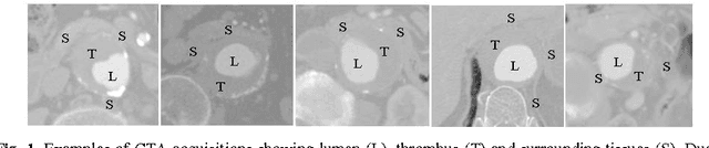


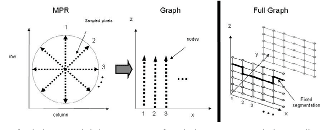
Simulation of arterial stenting procedures prior to intervention allows for appropriate device selection as well as highlights potential complications. To this end, we present a framework for facilitating virtual aortic stenting from a contrast computer tomography (CT) scan. More specifically, we present a method for both lumen and outer wall segmentation that may be employed in determining both the appropriateness of intervention as well as the selection and localization of the device. The more challenging recovery of the outer wall is based on a novel minimal closure tracking algorithm. Our aortic segmentation method has been validated on over 3000 multiplanar reformatting (MPR) planes from 50 CT angiography data sets yielding a Dice Similarity Coefficient (DSC) of 90.67%.