Soham Basu
carps: A Framework for Comparing N Hyperparameter Optimizers on M Benchmarks
Jun 06, 2025


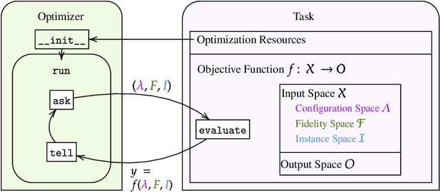
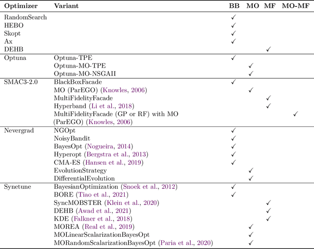
Abstract:Hyperparameter Optimization (HPO) is crucial to develop well-performing machine learning models. In order to ease prototyping and benchmarking of HPO methods, we propose carps, a benchmark framework for Comprehensive Automated Research Performance Studies allowing to evaluate N optimizers on M benchmark tasks. In this first release of carps, we focus on the four most important types of HPO task types: blackbox, multi-fidelity, multi-objective and multi-fidelity-multi-objective. With 3 336 tasks from 5 community benchmark collections and 28 variants of 9 optimizer families, we offer the biggest go-to library to date to evaluate and compare HPO methods. The carps framework relies on a purpose-built, lightweight interface, gluing together optimizers and benchmark tasks. It also features an analysis pipeline, facilitating the evaluation of optimizers on benchmarks. However, navigating a huge number of tasks while developing and comparing methods can be computationally infeasible. To address this, we obtain a subset of representative tasks by minimizing the star discrepancy of the subset, in the space spanned by the full set. As a result, we propose an initial subset of 10 to 30 diverse tasks for each task type, and include functionality to re-compute subsets as more benchmarks become available, enabling efficient evaluations. We also establish a first set of baseline results on these tasks as a measure for future comparisons. With carps (https://www.github.com/automl/CARP-S), we make an important step in the standardization of HPO evaluation.
A Comparative Study of Multiple Deep Learning Algorithms for Efficient Localization of Bone Joints in the Upper Limbs of Human Body
Oct 28, 2024Abstract:This paper addresses the medical imaging problem of joint detection in the upper limbs, viz. elbow, shoulder, wrist and finger joints. Localization of joints from X-Ray and Computerized Tomography (CT) scans is an essential step for the assessment of various bone-related medical conditions like Osteoarthritis, Rheumatoid Arthritis, and can even be used for automated bone fracture detection. Automated joint localization also detects the corresponding bones and can serve as input to deep learning-based models used for the computerized diagnosis of the aforementioned medical disorders. This in-creases the accuracy of prediction and aids the radiologists with analyzing the scans, which is quite a complex and exhausting task. This paper provides a detailed comparative study between diverse Deep Learning (DL) models - YOLOv3, YOLOv7, EfficientDet and CenterNet in multiple bone joint detections in the upper limbs of the human body. The research analyses the performance of different DL models, mathematically, graphically and visually. These models are trained and tested on a portion of the openly available MURA (musculoskeletal radiographs) dataset. The study found that the best Mean Average Precision (mAP at 0.5:0.95) values of YOLOv3, YOLOv7, EfficientDet and CenterNet are 35.3, 48.3, 46.5 and 45.9 respectively. Besides, it has been found YOLOv7 performed the best for accurately predicting the bounding boxes while YOLOv3 performed the worst in the Visual Analysis test. Code available at https://github.com/Sohambasu07/BoneJointsLocalization
Segmentation of Blood Vessels, Optic Disc Localization, Detection of Exudates and Diabetic Retinopathy Diagnosis from Digital Fundus Images
Jul 09, 2022Abstract:Diabetic Retinopathy (DR) is a complication of long-standing, unchecked diabetes and one of the leading causes of blindness in the world. This paper focuses on improved and robust methods to extract some of the features of DR, viz. Blood Vessels and Exudates. Blood vessels are segmented using multiple morphological and thresholding operations. For the segmentation of exudates, k-means clustering and contour detection on the original images are used. Extensive noise reduction is performed to remove false positives from the vessel segmentation algorithm's results. The localization of Optic Disc using k-means clustering and template matching is also performed. Lastly, this paper presents a Deep Convolutional Neural Network (DCNN) model with 14 Convolutional Layers and 2 Fully Connected Layers, for the automatic, binary diagnosis of DR. The vessel segmentation, optic disc localization and DCNN achieve accuracies of 95.93%, 98.77% and 75.73% respectively. The source code and pre-trained model are available https://github.com/Sohambasu07/DR_2021
* RAAI 2020, 11 pages, 12 figures, 2 tables
 Add to Chrome
 Add to Firefox
 Add to Edge