Smriti Regmi
Classification of Endoscopy and Video Capsule Images using CNN-Transformer Model
Aug 20, 2024


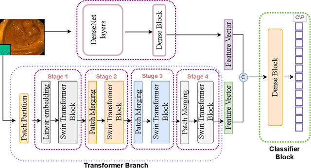
Abstract:Gastrointestinal cancer is a leading cause of cancer-related incidence and death, making it crucial to develop novel computer-aided diagnosis systems for early detection and enhanced treatment. Traditional approaches rely on the expertise of gastroenterologists to identify diseases; however, this process is subjective, and interpretation can vary even among expert clinicians. Considering recent advancements in classifying gastrointestinal anomalies and landmarks in endoscopic and video capsule endoscopy images, this study proposes a hybrid model that combines the advantages of Transformers and Convolutional Neural Networks (CNNs) to enhance classification performance. Our model utilizes DenseNet201 as a CNN branch to extract local features and integrates a Swin Transformer branch for global feature understanding, combining both to perform the classification task. For the GastroVision dataset, our proposed model demonstrates excellent performance with Precision, Recall, F1 score, Accuracy, and Matthews Correlation Coefficient (MCC) of 0.8320, 0.8386, 0.8324, 0.8386, and 0.8191, respectively, showcasing its robustness against class imbalance and surpassing other CNNs as well as the Swin Transformer model. Similarly, for the Kvasir-Capsule, a large video capsule endoscopy dataset, our model outperforms all others, achieving overall Precision, Recall, F1 score, Accuracy, and MCC of 0.7007, 0.7239, 0.6900, 0.7239, and 0.3871. Moreover, we generated saliency maps to explain our model's focus areas, demonstrating its reliable decision-making process. The results underscore the potential of our hybrid CNN-Transformer model in aiding the early and accurate detection of gastrointestinal (GI) anomalies.
Vision Transformer for Efficient Chest X-ray and Gastrointestinal Image Classification
Apr 23, 2023


Abstract:Medical image analysis is a hot research topic because of its usefulness in different clinical applications, such as early disease diagnosis and treatment. Convolutional neural networks (CNNs) have become the de-facto standard in medical image analysis tasks because of their ability to learn complex features from the available datasets, which makes them surpass humans in many image-understanding tasks. In addition to CNNs, transformer architectures also have gained popularity for medical image analysis tasks. However, despite progress in the field, there are still potential areas for improvement. This study uses different CNNs and transformer-based methods with a wide range of data augmentation techniques. We evaluated their performance on three medical image datasets from different modalities. We evaluated and compared the performance of the vision transformer model with other state-of-the-art (SOTA) pre-trained CNN networks. For Chest X-ray, our vision transformer model achieved the highest F1 score of 0.9532, recall of 0.9533, Matthews correlation coefficient (MCC) of 0.9259, and ROC-AUC score of 0.97. Similarly, for the Kvasir dataset, we achieved an F1 score of 0.9436, recall of 0.9437, MCC of 0.9360, and ROC-AUC score of 0.97. For the Kvasir-Capsule (a large-scale VCE dataset), our ViT model achieved a weighted F1-score of 0.7156, recall of 0.7182, MCC of 0.3705, and ROC-AUC score of 0.57. We found that our transformer-based models were better or more effective than various CNN models for classifying different anatomical structures, findings, and abnormalities. Our model showed improvement over the CNN-based approaches and suggests that it could be used as a new benchmarking algorithm for algorithm development.
 Add to Chrome
 Add to Firefox
 Add to Edge