Sergio Garcia-Garcia
Enhanced Mortality Prediction In Patients With Subarachnoid Haemorrhage Using A Deep Learning Model Based On The Initial CT Scan
Aug 25, 2023


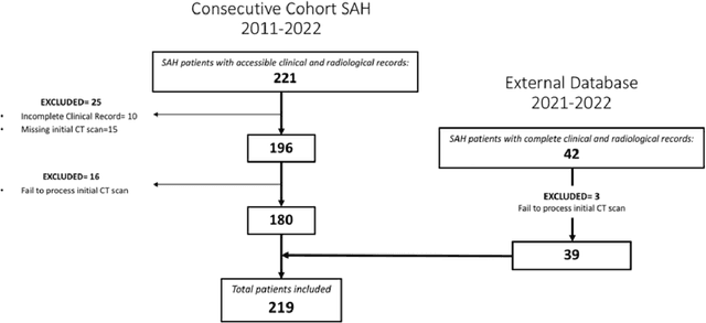
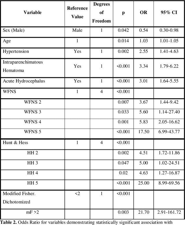
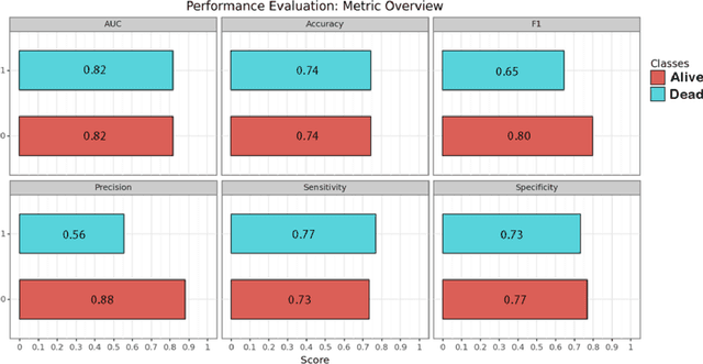
Abstract:PURPOSE: Subarachnoid hemorrhage (SAH) entails high morbidity and mortality rates. Convolutional neural networks (CNN), a form of deep learning, are capable of generating highly accurate predictions from imaging data. Our objective was to predict mortality in SAH patients by processing the initial CT scan on a CNN based algorithm. METHODS: Retrospective multicentric study of a consecutive cohort of patients with SAH between 2011-2022. Demographic, clinical and radiological variables were analyzed. Pre-processed baseline CT scan images were used as the input for training a CNN using AUCMEDI Framework. Our model's architecture leverages the DenseNet-121 structure, employing transfer learning principles. The output variable was mortality in the first three months. Performance of the model was evaluated by statistical parameters conventionally used in studies involving artificial intelligence methods. RESULTS: Images from 219 patients were processed, 175 for training and validation of the CNN and 44 for its evaluation. 52%(115/219) of patients were female, and the median age was 58(SD=13.06) years. 18.5%(39/219) were idiopathic SAH. Mortality rate was 28.5%(63/219). The model showed good accuracy at predicting mortality in SAH patients exclusively using the images of the initial CT scan (Accuracy=74%, F1=75% and AUC=82%). CONCLUSION: Modern image processing techniques based on AI and CNN make possible to predict mortality in SAH patients with high accuracy using CT scan images as the only input. These models might be optimized by including more data and patients resulting in better training, development and performance on tasks which are beyond the skills of conventional clinical knowledge.
The Rio Hortega University Hospital Glioblastoma dataset: a comprehensive collection of preoperative, early postoperative and recurrence MRI scans (RHUH-GBM)
May 02, 2023
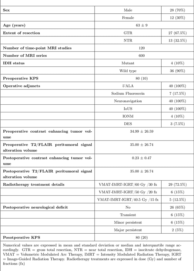
Abstract:Glioblastoma, a highly aggressive primary brain tumor, is associated with poor patient outcomes. Although magnetic resonance imaging (MRI) plays a critical role in diagnosing, characterizing, and forecasting glioblastoma progression, public MRI repositories present significant drawbacks, including insufficient postoperative and follow-up studies as well as expert tumor segmentations. To address these issues, we present the "R\'io Hortega University Hospital Glioblastoma Dataset (RHUH-GBM)," a collection of multiparametric MRI images, volumetric assessments, molecular data, and survival details for glioblastoma patients who underwent total or near-total enhancing tumor resection. The dataset features expert-corrected segmentations of tumor subregions, offering valuable ground truth data for developing algorithms for postoperative and follow-up MRI scans. The public release of the RHUH-GBM dataset significantly contributes to glioblastoma research, enabling the scientific community to study recurrence patterns and develop new diagnostic and prognostic models. This may result in more personalized, effective treatments and ultimately improved patient outcomes.
 Add to Chrome
 Add to Firefox
 Add to Edge