Naira Elazab
Breast Cancer Classification Based on Histopathological Images Using a Deep Learning Capsule Network
Aug 01, 2022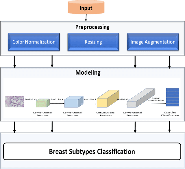
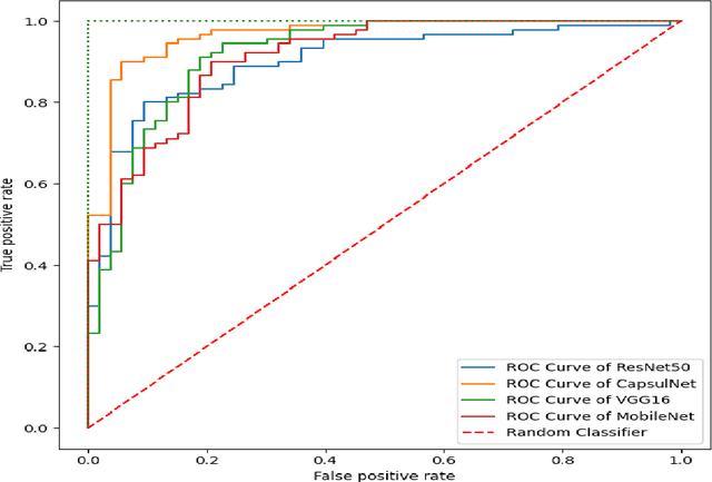
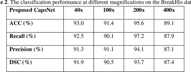
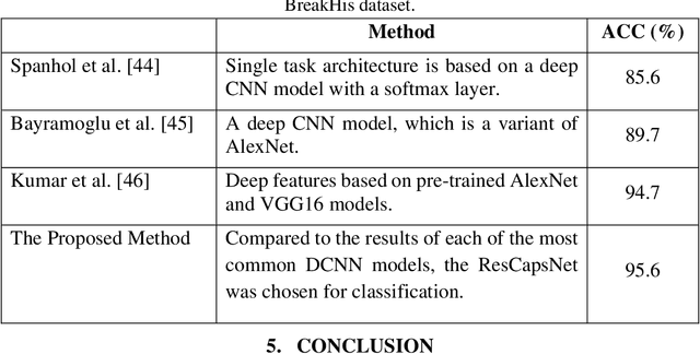
Abstract:Breast cancer is one of the most serious types of cancer that can occur in women. The automatic diagnosis of breast cancer by analyzing histological images (HIs) is important for patients and their prognosis. The classification of HIs provides clinicians with an accurate understanding of diseases and allows them to treat patients more efficiently. Deep learning (DL) approaches have been successfully employed in a variety of fields, particularly medical imaging, due to their capacity to extract features automatically. This study aims to classify different types of breast cancer using HIs. In this research, we present an enhanced capsule network that extracts multi-scale features using the Res2Net block and four additional convolutional layers. Furthermore, the proposed method has fewer parameters due to using small convolutional kernels and the Res2Net block. As a result, the new method outperforms the old ones since it automatically learns the best possible features. The testing results show that the model outperformed the previous DL methods.
Objective Diagnosis for Histopathological Images Based on Machine Learning Techniques: Classical Approaches and New Trends
Nov 10, 2020
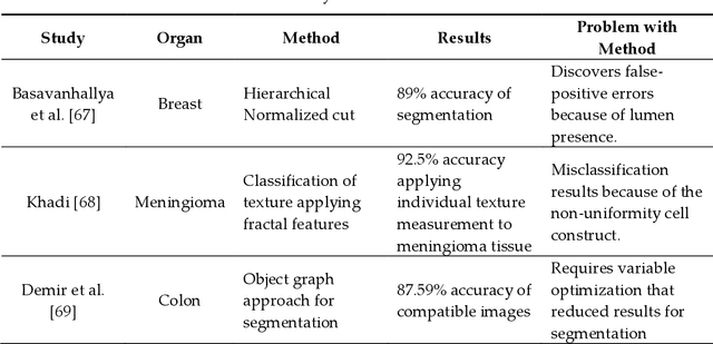

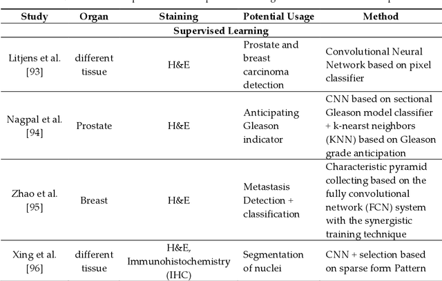
Abstract:Histopathology refers to the examination by a pathologist of biopsy samples. Histopathology images are captured by a microscope to locate, examine, and classify many diseases, such as different cancer types. They provide a detailed view of different types of diseases and their tissue status. These images are an essential resource with which to define biological compositions or analyze cell and tissue structures. This imaging modality is very important for diagnostic applications. The analysis of histopathology images is a prolific and relevant research area supporting disease diagnosis. In this paper, the challenges of histopathology image analysis are evaluated. An extensive review of conventional and deep learning techniques which have been applied in histological image analyses is presented. This review summarizes many current datasets and highlights important challenges and constraints with recent deep learning techniques, alongside possible future research avenues. Despite the progress made in this research area so far, it is still a significant area of open research because of the variety of imaging techniques and disease-specific characteristics.
* 26 Pages, 5 figures, 4 tables
 Add to Chrome
 Add to Firefox
 Add to Edge