Ahmed Elgarayhi
Efficient liver segmentation with 3D CNN using computed tomography scans
Aug 28, 2022Abstract:The liver is one of the most critical metabolic organs in vertebrates due to its vital functions in the human body, such as detoxification of the blood from waste products and medications. Liver diseases due to liver tumors are one of the most common mortality reasons around the globe. Hence, detecting liver tumors in the early stages of tumor development is highly required as a critical part of medical treatment. Many imaging modalities can be used as aiding tools to detect liver tumors. Computed tomography (CT) is the most used imaging modality for soft tissue organs such as the liver. This is because it is an invasive modality that can be captured relatively quickly. This paper proposed an efficient automatic liver segmentation framework to detect and segment the liver out of CT abdomen scans using the 3D CNN DeepMedic network model. Segmenting the liver region accurately and then using the segmented liver region as input to tumors segmentation method is adopted by many studies as it reduces the false rates resulted from segmenting abdomen organs as tumors. The proposed 3D CNN DeepMedic model has two pathways of input rather than one pathway, as in the original 3D CNN model. In this paper, the network was supplied with multiple abdomen CT versions, which helped improve the segmentation quality. The proposed model achieved 94.36%, 94.57%, 91.86%, and 93.14% for accuracy, sensitivity, specificity, and Dice similarity score, respectively. The experimental results indicate the applicability of the proposed method.
Breast Cancer Classification Based on Histopathological Images Using a Deep Learning Capsule Network
Aug 01, 2022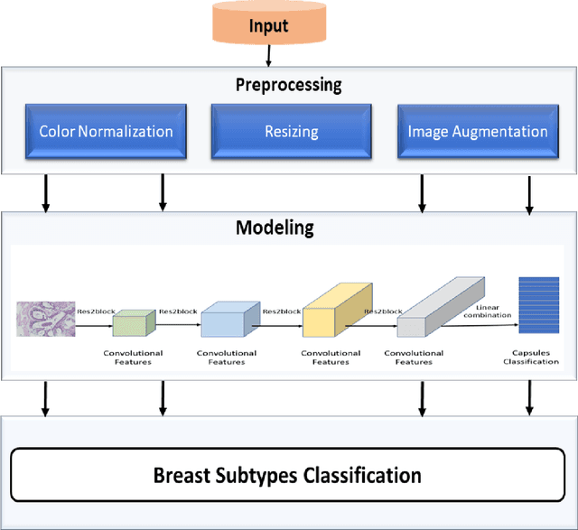
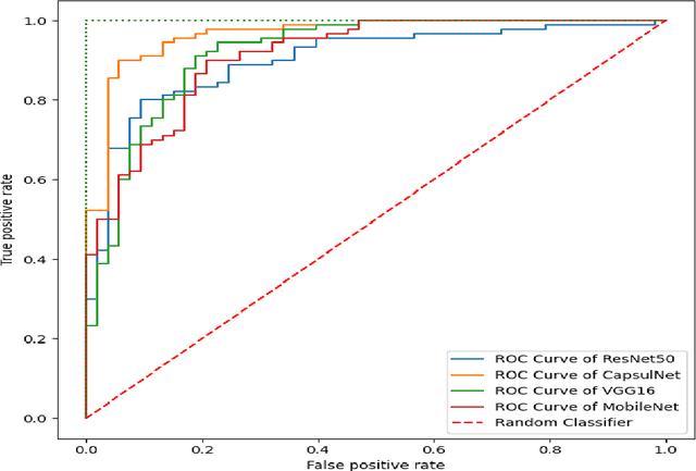
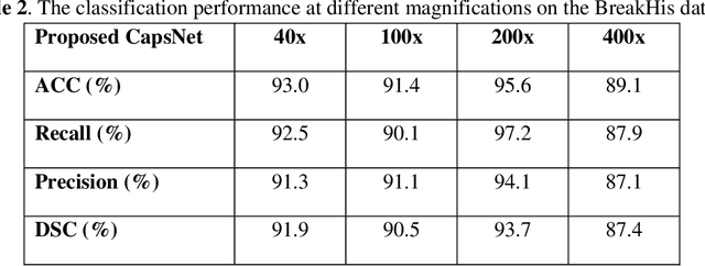
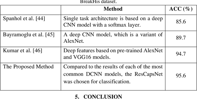
Abstract:Breast cancer is one of the most serious types of cancer that can occur in women. The automatic diagnosis of breast cancer by analyzing histological images (HIs) is important for patients and their prognosis. The classification of HIs provides clinicians with an accurate understanding of diseases and allows them to treat patients more efficiently. Deep learning (DL) approaches have been successfully employed in a variety of fields, particularly medical imaging, due to their capacity to extract features automatically. This study aims to classify different types of breast cancer using HIs. In this research, we present an enhanced capsule network that extracts multi-scale features using the Res2Net block and four additional convolutional layers. Furthermore, the proposed method has fewer parameters due to using small convolutional kernels and the Res2Net block. As a result, the new method outperforms the old ones since it automatically learns the best possible features. The testing results show that the model outperformed the previous DL methods.
An Enhanced Deep Learning Technique for Prostate Cancer Identification Based on MRI Scans
Aug 01, 2022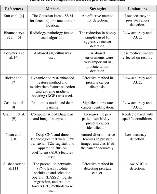


Abstract:Prostate cancer is the most dangerous cancer diagnosed in men worldwide. Prostate diagnosis has been affected by many factors, such as lesion complexity, observer visibility, and variability. Many techniques based on Magnetic Resonance Imaging (MRI) have been used for prostate cancer identification and classification in the last few decades. Developing these techniques is crucial and has a great medical effect because they improve the treatment benefits and the chance of patients' survival. A new technique that depends on MRI has been proposed to improve the diagnosis. This technique consists of two stages. First, the MRI images have been preprocessed to make the medical image more suitable for the detection step. Second, prostate cancer identification has been performed based on a pre-trained deep learning model, InceptionResNetV2, that has many advantages and achieves effective results. In this paper, the InceptionResNetV2 deep learning model used for this purpose has average accuracy equals to 89.20%, and the area under the curve (AUC) equals to 93.6%. The experimental results of this proposed new deep learning technique represent promising and effective results compared to other previous techniques.
 Add to Chrome
 Add to Firefox
 Add to Edge