Melanie Rieff
SMMILE: An Expert-Driven Benchmark for Multimodal Medical In-Context Learning
Jun 26, 2025


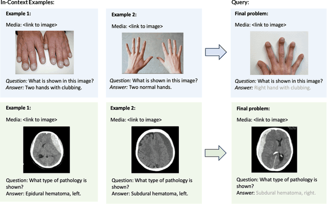
Abstract:Multimodal in-context learning (ICL) remains underexplored despite significant potential for domains such as medicine. Clinicians routinely encounter diverse, specialized tasks requiring adaptation from limited examples, such as drawing insights from a few relevant prior cases or considering a constrained set of differential diagnoses. While multimodal large language models (MLLMs) have shown advances in medical visual question answering (VQA), their ability to learn multimodal tasks from context is largely unknown. We introduce SMMILE, the first expert-driven multimodal ICL benchmark for medical tasks. Eleven medical experts curated problems, each including a multimodal query and multimodal in-context examples as task demonstrations. SMMILE encompasses 111 problems (517 question-image-answer triplets) covering 6 medical specialties and 13 imaging modalities. We further introduce SMMILE++, an augmented variant with 1038 permuted problems. A comprehensive evaluation of 15 MLLMs demonstrates that most models exhibit moderate to poor multimodal ICL ability in medical tasks. In open-ended evaluations, ICL contributes only 8% average improvement over zero-shot on SMMILE and 9.4% on SMMILE++. We observe a susceptibility for irrelevant in-context examples: even a single noisy or irrelevant example can degrade performance by up to 9.5%. Moreover, example ordering exhibits a recency bias, i.e., placing the most relevant example last can lead to substantial performance improvements by up to 71%. Our findings highlight critical limitations and biases in current MLLMs when learning multimodal medical tasks from context.
U-net based prediction of cerebrospinal fluid distribution and ventricular reflux grading
Oct 06, 2024


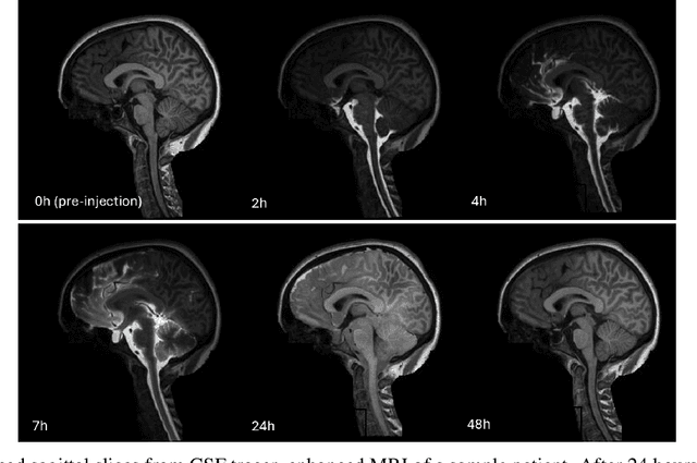
Abstract:Previous work shows evidence that cerebrospinal fluid (CSF) plays a crucial role in brain waste clearance processes, and that altered flow patterns are associated with various diseases of the central nervous system. In this study, we investigate the potential of deep learning to predict the distribution in human brain of a gadolinium-based CSF contrast agent (tracer) administered intrathecal. For this, T1-weighted magnetic resonance imaging (MRI) scans taken at multiple time points before and after intrathecal injection were utilized. We propose a U-net-based supervised learning model to predict pixel-wise signal increases at their peak after 24 hours. Its performance is evaluated based on different tracer distribution stages provided during training, including predictions from baseline scans taken before injection. Our findings indicate that using imaging data from just the first two hours post-injection for training yields tracer flow predictions comparable to those trained with additional later-stage scans. The model was further validated by comparing ventricular reflux gradings provided by neuroradiologists, and inter-rater grading among medical experts and the model showed excellent agreement. Our results demonstrate the potential of deep learning-based methods for CSF flow prediction, suggesting that fewer MRI scans could be sufficient for clinical analysis, which might significantly improve clinical efficiency, patient well-being, and lower healthcare costs.
 Add to Chrome
 Add to Firefox
 Add to Edge