Martina Paccini
Framework of a multiscale data-driven digital twin of the muscle-skeletal system
Jun 13, 2025Abstract:Musculoskeletal disorders (MSDs) are a leading cause of disability worldwide, requiring advanced diagnostic and therapeutic tools for personalised assessment and treatment. Effective management of MSDs involves the interaction of heterogeneous data sources, making the Digital Twin (DT) paradigm a valuable option. This paper introduces the Musculoskeletal Digital Twin (MS-DT), a novel framework that integrates multiscale biomechanical data with computational modelling to create a detailed, patient-specific representation of the musculoskeletal system. By combining motion capture, ultrasound imaging, electromyography, and medical imaging, the MS-DT enables the analysis of spinal kinematics, posture, and muscle function. An interactive visualisation platform provides clinicians and researchers with an intuitive interface for exploring biomechanical parameters and tracking patient-specific changes. Results demonstrate the effectiveness of MS-DT in extracting precise kinematic and dynamic tissue features, offering a comprehensive tool for monitoring spine biomechanics and rehabilitation. This framework provides high-fidelity modelling and real-time visualization to improve patient-specific diagnosis and intervention planning.
3D Skin Segmentation Methods in Medical Imaging: A Comparison
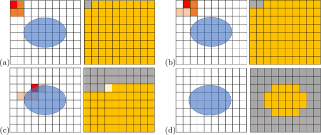
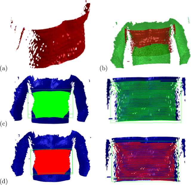
Jun 13, 2025Abstract:Automatic segmentation of anatomical structures is critical in medical image analysis, aiding diagnostics and treatment planning. Skin segmentation plays a key role in registering and visualising multimodal imaging data. 3D skin segmentation enables applications in personalised medicine, surgical planning, and remote monitoring, offering realistic patient models for treatment simulation, procedural visualisation, and continuous condition tracking. This paper analyses and compares algorithmic and AI-driven skin segmentation approaches, emphasising key factors to consider when selecting a strategy based on data availability and application requirements. We evaluate an iterative region-growing algorithm and the TotalSegmentator, a deep learning-based approach, across different imaging modalities and anatomical regions. Our tests show that AI segmentation excels in automation but struggles with MRI due to its CT-based training, while the graphics-based method performs better for MRIs but introduces more noise. AI-driven segmentation also automates patient bed removal in CT, whereas the graphics-based method requires manual intervention.
US & MR Image-Fusion Based on Skin Co-Registration
Jul 26, 2023


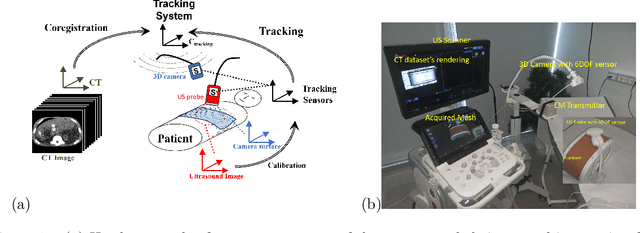
Abstract:The study and development of innovative solutions for the advanced visualisation, representation and analysis of medical images offer different research directions. Current practice in medical imaging consists in combining real-time US with imaging modalities that allow internal anatomy acquisitions, such as CT, MRI, PET or similar. Application of image-fusion approaches can be found in tracking surgical tools and/or needles, in real-time during interventions. Thus, this work proposes a fusion imaging system for the registration of CT and MRI images with real-time US acquisition leveraging a 3D camera sensor. The main focus of the work is the portability of the system and its applicability to different anatomical districts.
3D Patient-specific Modelling and Characterisation of Muscle-Skeletal Districts
Apr 18, 2023Abstract:This work addresses the patient-specific characterisation of the morphology and pathologies of muscle-skeletal districts (e.g., wrist, spine) to support diagnostic activities and follow-up exams through the integration of morphological and tissue information. We propose different methods for the integration of morphological information, retrieved from the geometrical analysis of 3D surface models, with tissue information extracted from volume images. For the qualitative and quantitative validation, we will discuss the localisation of bone erosion sites on the wrists to monitor rheumatic diseases and the characterisation of the three functional regions of the spinal vertebrae to study the presence of osteoporotic fractures. The proposed approach supports the quantitative and visual evaluation of possible damages, surgery planning, and early diagnosis or follow-up studies. Finally, our analysis is general enough to be applied to different districts.
 Add to Chrome
 Add to Firefox
 Add to Edge