Laura Beretta
Dimensionality Reduction and Nearest Neighbors for Improving Out-of-Distribution Detection in Medical Image Segmentation
Aug 05, 2024


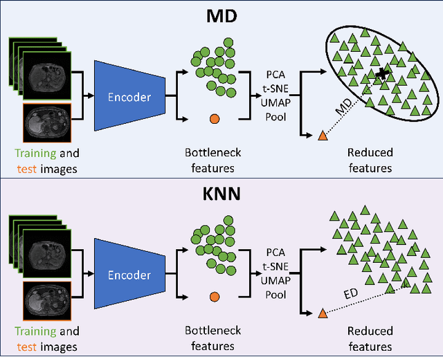
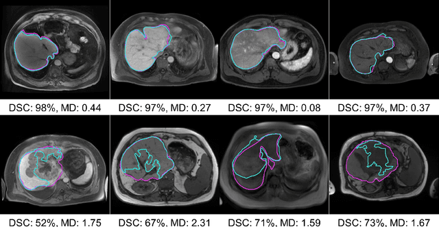
Abstract:Clinically deployed deep learning-based segmentation models are known to fail on data outside of their training distributions. While clinicians review the segmentations, these models tend to perform well in most instances, which could exacerbate automation bias. Therefore, detecting out-of-distribution images at inference is critical to warn the clinicians that the model likely failed. This work applied the Mahalanobis distance (MD) post hoc to the bottleneck features of four Swin UNETR and nnU-net models that segmented the liver on T1-weighted magnetic resonance imaging and computed tomography. By reducing the dimensions of the bottleneck features with either principal component analysis or uniform manifold approximation and projection, images the models failed on were detected with high performance and minimal computational load. In addition, this work explored a non-parametric alternative to the MD, a k-th nearest neighbors distance (KNN). KNN drastically improved scalability and performance over MD when both were applied to raw and average-pooled bottleneck features.
Heterogeneous Image-based Classification Using Distributional Data Analysis
Mar 11, 2024Abstract:Diagnostic imaging has gained prominence as potential biomarkers for early detection and diagnosis in a diverse array of disorders including cancer. However, existing methods routinely face challenges arising from various factors such as image heterogeneity. We develop a novel imaging-based distributional data analysis (DDA) approach that incorporates the probability (quantile) distribution of the pixel-level features as covariates. The proposed approach uses a smoothed quantile distribution (via a suitable basis representation) as functional predictors in a scalar-on-functional quantile regression model. Some distinctive features of the proposed approach include the ability to: (i) account for heterogeneity within the image; (ii) incorporate granular information spanning the entire distribution; and (iii) tackle variability in image sizes for unregistered images in cancer applications. Our primary goal is risk prediction in Hepatocellular carcinoma that is achieved via predicting the change in tumor grades at post-diagnostic visits using pre-diagnostic enhancement pattern mapping (EPM) images of the liver. Along the way, the proposed DDA approach is also used for case versus control diagnosis and risk stratification objectives. Our analysis reveals that when coupled with global structural radiomics features derived from the corresponding T1-MRI scans, the proposed smoothed quantile distributions derived from EPM images showed considerable improvements in sensitivity and comparable specificity in contrast to classification based on routinely used summary measures that do not account for image heterogeneity. Given that there are limited predictive modeling approaches based on heterogeneous images in cancer, the proposed method is expected to provide considerable advantages in image-based early detection and risk prediction.
 Add to Chrome
 Add to Firefox
 Add to Edge