 Add to Chrome
 Add to Firefox
 Add to Edge
CHMMOTv1 -- Cardiac and Hepatic Multi-Echo MRI Images and Clinical Dataset for Iron Overload on Thalassemia Patients
May 17, 2023Owing to the invasiveness and low accuracy of other tests, including biopsy and ferritin levels, magnetic resonance imaging (T2 and T2*-MRI) has been considered the standard test for patients with thalassemia (THM). Regarding deep learning networks in medical sciences for improving diagnosis and treatment purposes and the existence of minimal resources for them, we decided to provide a set of magnetic resonance images of the cardiac and hepatic organs. The dataset included 124 patients (67 women and 57 men) with a THM age range of (5-52) years. In addition, patients were divided into two groups: with follow-up (1-5 times) at time intervals of about (5-6) months and without follow-up. Also, T2* and, R2* values, the results of the cardiac and hepatic report (normal, mild, moderate, severe, and very severe), and laboratory tests including Ferritin, Bilirubin (D, and T), AST, ALT, and ALP levels were provided as an Excel file. This dataset CHMMOTv1) has been published in Mendeley Dataverse and is accessible through the web at: http://databiox.com.
Attention U-net approach in predicting Intensity Modulated Radiation Therapy dose distribution in brain glioma tumor
May 10, 2023


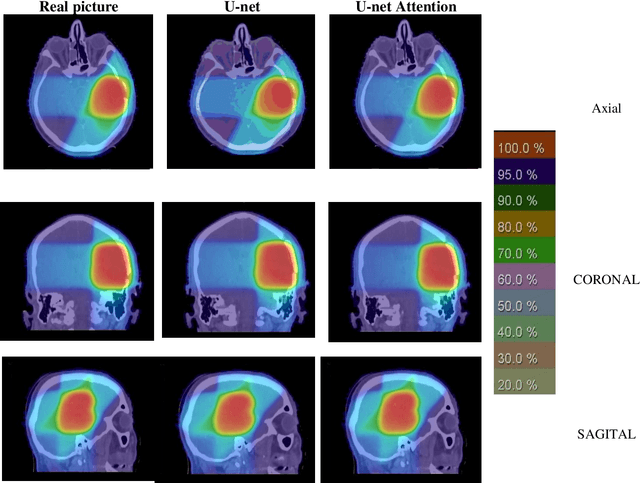
Today, intensity-modulated radiation therapy (IMRT) is one of the methods used to treat brain tumors. In conventional treatment planning methods, after identifying planning target volume (PTV), and organs at risk (OARs), and determining the limitations for them to receive radiation, the dose distribution is performed based on optimization algorithms, which is usually a time-consuming method. In this article, artificial intelligence is used to acquire the knowledge used in the treatment planning of past patients and to plan for new patients to speed up the process of treatment planning and determination of the appropriate dose distribution. In this paper, using deep learning algorithms, two different approaches are studied to predict dose distribution and compared with actual dose distributions. In the first method, only the images containing PTV and the distribution of the corresponding doses are used to train the convolutional neural network, but in the second one, in addition to PTV, the contours of four OARs are also used to introduce the network. The results show that the performance of both methods on test patients have high accuracy and in comparison with each other almost have the same results and high speed to design the dose distribution. Because the Only-PTV method does not have the process of OARs identifying, applying it in designing the dose distribution will be much faster than using the PTV-OARs method in the whole of treatment planning.
A High-Resolution Chest CT-Scan Image Dataset for COVID-19 Diagnosis and Differentiation
May 06, 2022
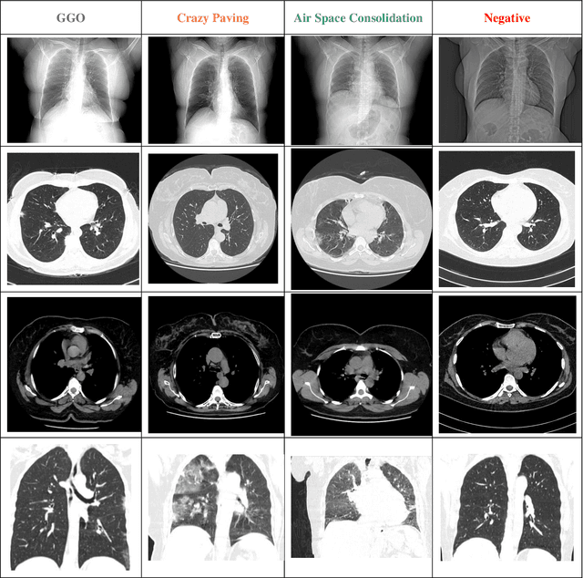
During the COVID-19 pandemic, computed tomography (CT) is a good way to diagnose COVID-19 patients. HRCT (High-Resolution Computed Tomography) is a form of computed tomography that uses advanced methods to improve image resolution. Publicly accessible COVID-19 CT image datasets are very difficult to come by due to privacy concerns, which impedes the study and development of AI-powered COVID-19 diagnostic algorithms based on CT images. To address this problem, we have introduced HRCTv1-COVID-19, a new COVID-19 high resolution chest CT Scan image dataset that includes not only COVID-19 cases of Ground Glass Opacity (GGO), Crazy Paving, and Air Space Consolidation, but also CT images of cases with negative COVID-19. The HRCTv1-COVID-19 dataset, which includes slice-level, and patient-level labels, has the potential to aid COVID-19 research, especially for diagnosis and differentiation using artificial intelligence algorithms, machine learning and deep learning methods. This dataset is accessible through web at: http://databiox.com and includes 181,106 chest HRCT images from 395 patients with four labels: GGO, Crazy Paving, Air Space Consolidation and Negative. Keywords- Dataset, COVID-19, CT-Scan, Computed Tomography, Medical Imaging, Chest Image.