 Add to Chrome
 Add to Firefox
 Add to Edge
Translation of Fetal Brain Ultrasound Images into Pseudo-MRI Images using Artificial Intelligence
Apr 03, 2025Ultrasound is a widely accessible and cost-effective medical imaging tool commonly used for prenatal evaluation of the fetal brain. However, it has limitations, particularly in the third trimester, where the complexity of the fetal brain requires high image quality for extracting quantitative data. In contrast, magnetic resonance imaging (MRI) offers superior image quality and tissue differentiation but is less available, expensive, and requires time-consuming acquisition. Thus, transforming ultrasonic images into an MRI-mimicking display may be advantageous and allow better tissue anatomy presentation. To address this goal, we have examined the use of artificial intelligence, implementing a diffusion model renowned for generating high-quality images. The proposed method, termed "Dual Diffusion Imposed Correlation" (DDIC), leverages a diffusion-based translation methodology, assuming a shared latent space between ultrasound and MRI domains. Model training was obtained utilizing the "HC18" dataset for ultrasound and the "CRL fetal brain atlas" along with the "FeTA " datasets for MRI. The generated pseudo-MRI images provide notable improvements in visual discrimination of brain tissue, especially in the lateral ventricles and the Sylvian fissure, characterized by enhanced contrast clarity. Improvement was demonstrated in Mutual information, Peak signal-to-noise ratio, Fr\'echet Inception Distance, and Contrast-to-noise ratio. Findings from these evaluations indicate statistically significant superior performance of the DDIC compared to other translation methodologies. In addition, a Medical Opinion Test was obtained from 5 gynecologists. The results demonstrated display improvement in 81% of the tested images. In conclusion, the presented pseudo-MRI images hold the potential for streamlining diagnosis and enhancing clinical outcomes through improved representation.
Image translation of Ultrasound to Pseudo Anatomical Display Using Artificial Intelligence
Feb 16, 2022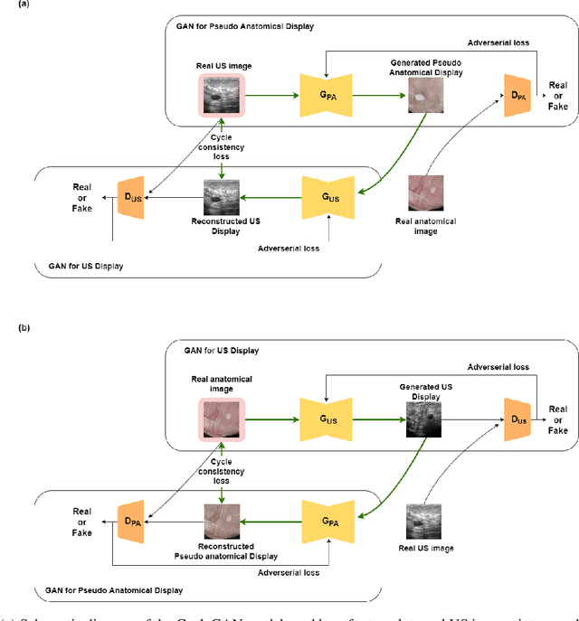
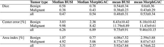
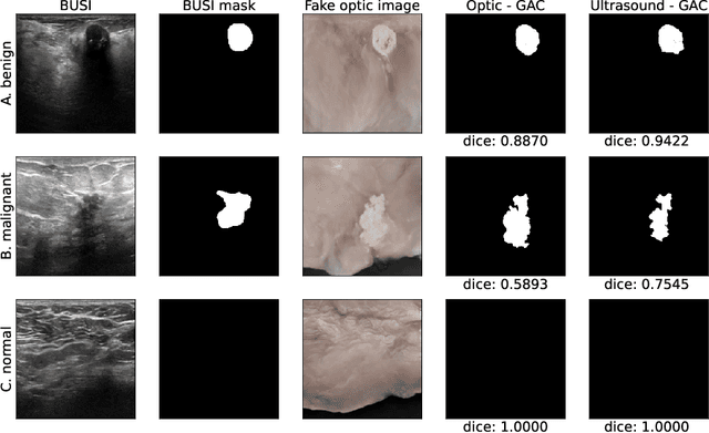
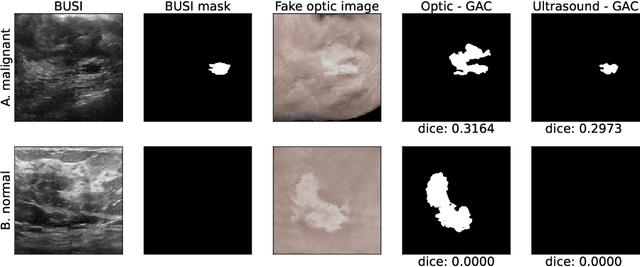
Ultrasound is the second most used modality in medical imaging. It is cost effective, hazardless, portable and implemented routinely in numerous clinical procedures. Nonetheless, image quality is characterized by granulated appearance, poor SNR and speckle noise. Specific for malignant tumors, the margins are blurred and indistinct. Thus, there is a great need for improving ultrasound image quality. We hypothesize that this can be achieved by translation into a more realistic anatomic display, using neural networks. In order to achieve this goal, the preferable approach would be to use a set of paired images. However, this is practically impossible in our case. Therefore, CycleGAN was used, to learn each domain properties separately and enforce cross domain cycle consistency. The two datasets which were used for training the model were "Breast Ultrasound Images" (BUSI) and a set of optic images of poultry breast tissue samples acquired at our lab. The generated pseudo anatomical images provide improved visual discrimination of the lesions with clearer border definition and pronounced contrast. Furthermore, the algorithm manages to overcome the acoustic shadows artifacts commonly appearing in ultrasonic images. In order to evaluate the preservation of the anatomical features, the lesions in the ultrasonic images and the generated pseudo anatomical images were both automatically segmented and compared. This comparison yielded median dice score of 0.78 for the benign tumors and 0.43 for the malignancies. Median lesion center error of 2.38% and 8.42% for the benign and malignancies respectively and median area error index of 0.77% and 5.06% for the benign and malignancies respectively. In conclusion, these generated pseudo anatomical images, which are presented in a more intuitive way, preserve tissue anatomy and can potentially simplify the diagnosis and improve the clinical outcome.