Gabriele Multhoff
Eigenspectra optoacoustic tomography achieves quantitative blood oxygenation imaging deep in tissues
Nov 18, 2015


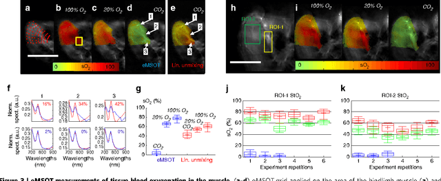
Abstract:Light propagating in tissue attains a spectrum that varies with location due to wavelength-dependent fluence attenuation by tissue optical properties, an effect that causes spectral corruption. Predictions of the spectral variations of light fluence in tissue are challenging since the spatial distribution of optical properties in tissue cannot be resolved in high resolution or with high accuracy by current methods. Spectral corruption has fundamentally limited the quantification accuracy of optical and optoacoustic methods and impeded the long sought-after goal of imaging blood oxygen saturation (sO2) deep in tissues; a critical but still unattainable target for the assessment of oxygenation in physiological processes and disease. We discover a new principle underlying light fluence in tissues, which describes the wavelength dependence of light fluence as an affine function of a few reference base spectra, independently of the specific distribution of tissue optical properties. This finding enables the introduction of a previously undocumented concept termed eigenspectra Multispectral Optoacoustic Tomography (eMSOT) that can effectively account for wavelength dependent light attenuation without explicit knowledge of the tissue optical properties. We validate eMSOT in more than 2000 simulations and with phantom and animal measurements. We find that eMSOT can quantitatively image tissue sO2 reaching in many occasions a better than 10-fold improved accuracy over conventional spectral optoacoustic methods. Then, we show that eMSOT can spatially resolve sO2 in muscle and tumor; revealing so far unattainable tissue physiology patterns. Last, we related eMSOT readings to cancer hypoxia and found congruence between eMSOT tumor sO2 images and tissue perfusion and hypoxia maps obtained by correlative histological analysis.
 Add to Chrome
 Add to Firefox
 Add to Edge