Debjani Paul
Single Test Image-Based Automated Machine Learning System for Distinguishing between Trait and Diseased Blood Samples
Mar 30, 2021


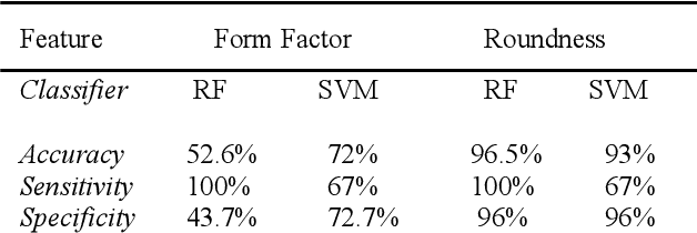
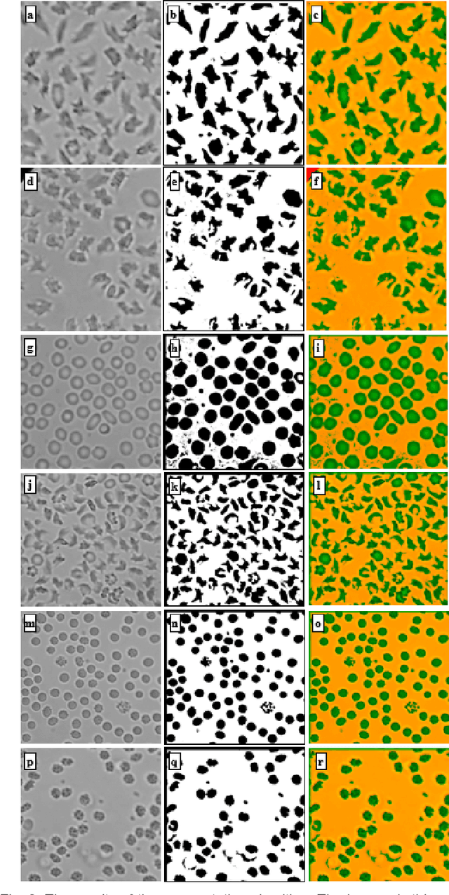
Abstract:We introduce a machine learning-based method for fully automated diagnosis of sickle cell disease of poor-quality unstained images of a mobile microscope. Our method is capable of distinguishing between diseased, trait (carrier), and normal samples unlike the previous methods that are limited to distinguishing the normal from the abnormal samples only. The novelty of this method comes from distinguishing the trait and the diseased samples from challenging images that have been captured directly in the field. The proposed approach contains two parts, the segmentation part followed by the classification part. We use a random forest algorithm to segment such challenging images acquitted through a mobile phone-based microscope. Then, we train two classifiers based on a random forest (RF) and a support vector machine (SVM) for classification. The results show superior performances of both of the classifiers not only for images which have been captured in the lab, but also for the ones which have been acquired in the field itself.
 Add to Chrome
 Add to Firefox
 Add to Edge